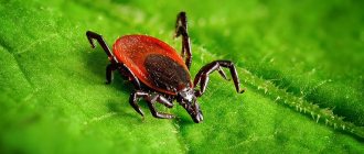
The tick is a common parasite whose activity period occurs in spring, summer and autumn. Throughout the warm period and even in the autumn cold, this insect looks for victims, waiting on blades of grass and bushes.
Even people in full clothing and animals treated with special means are not completely protected from tick bites. Therefore, you need to be prepared for the parasite to stick to the skin: know the likely consequences, the algorithm of necessary actions, and how to remove the tick.
What to do if the tick head remains in the human body
Removing a tick from under the skin Studying the tick and its particles What to do if the head of the tick remains in the human body, there are several options for behavior - remove it yourself, seek help from specialists, treat it with medicine, wait until it comes out on its own. You can carefully perform the operation at home, but if the wound has managed to fester, it is better to go to the clinic.
What happens if the tick's head remains in the human body?
There are several opinions on this matter. Some people are sure that there is nothing wrong. After a few days, it will be rejected by the body on its own. Another part of the victims insists that if the tick head remains inside the wound, it must be removed. In the future, this situation is dangerous for the development of a purulent process and infection.
How to pull out the proboscis
After unsuccessful extraction of the parasite, a head or proboscis may remain under the skin. If a black dot remains at the site of the bite, we are talking about the proboscis. To get rid of it, you can do the same as with an ordinary splinter.
- Treat the skin around with medical alcohol or any alcohol tincture. Disinfect the sharp needle, carefully pick it up, and pull out the tick sting.
- Lubricate the location of the body part and head with iodine. After a couple of days, the tick's proboscis will come out on its own.
How to remove a tick's head if it comes off
To remove the attached parasite, it is recommended to lubricate the surrounding skin with medical alcohol. After a few minutes, pick it up at the base with tweezers and twist it counterclockwise or clockwise. After a few turns the parasite will be outside. However, there is often a situation when part of the tick remains in the body.
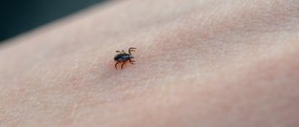
You can determine the presence of a tick head at the site of the bite either immediately by carefully examining the parasite, or you can understand it after a few days when swelling and redness appear. There is a depression in the center, a black dot. Below is a photo of a similar situation.
There are several ways to get the head of a tick.
- Wipe the bite site with a cotton swab soaked in alcohol to clearly see the location of the head. Disinfect a sharp needle or heat it over a burning candle. Pick up a piece of the parasite and pull it out like a splinter. Treat the skin with iodine and brilliant green.
- If the head and part of the body remain under the skin, remove it using forceps or tweezers. Clamp at the base and gently twist in any direction. The basic rule for a successful operation is not to change the direction of rotation, not to make sudden movements, and not to rush.
- Fold the gauze in several layers, apply Vishnevsky ointment liberally, apply to the sore spot, secure, and leave overnight. The next day, a piece of the parasite's body will come out along with the accumulated pus.
If independent actions do not give the desired result, you need to seek help from specialists. Under sterile conditions, the doctor makes an incision and removes the foreign body.
Is it possible to submit your head for analysis?
Ticks are carriers of dangerous diseases - borreliosis, tick-borne encephalitis. The first virus enters the human body with saliva during a bite, the second infection enters along with the blood of the infected parasite. This happens when the pest is removed incorrectly, the head is torn off, and the body may burst.
To be completely sure that there is no infection, the body of the extracted tick must be placed in a jar, with damp cotton wool placed at the bottom, and taken to the laboratory. In a few days, experts will render a verdict - whether the tick is infectious or not. Otherwise, you can find out about the presence of the disease within 14-30 days. This is how long the incubation period lasts. There is no point in taking tests before this time, they will not correspond to reality. For prevention after a bite, experts recommend vaccination or administration of immunoglobulin.
A live or dead tick should be brought to the laboratory. The main requirement is that it must be whole. The parasite must be placed in a humid environment, after moistening the cotton wool. There is no point in taking the body or the extracted head for analysis.
(1 rating, average 5 out of 5)
Is it possible to send a tick for examination in parts?
It is impossible to determine by the appearance of a tick whether it is infected or not. Despite the fact that not every individual carries pathogens of infectious diseases, it is better not to risk it and send the extracted parasite to the laboratory. The best option is if the tick was pulled out alive. Then the study will show the most accurate picture. If the bloodsucker died during the extraction or delivery process, it can also be tested, but this must be done as quickly as possible.
But it happens that the head of the parasite comes off when it is taken out. Worried about their health, some are trying to donate the body and severed head of the bloodsucker for research. But laboratories do not accept such material. Since if the tick was left without a head during the process of removal from the skin, then there is no point in doing tests, they will not give the correct result.
First aid for a tick bite
If a person was bitten far from a large populated area and it is not possible to go to a medical facility, you need to know what to do after a tick bite. It must be removed immediately upon discovery.
People prone to allergic reactions are recommended to take antihistamine tablets when bitten by a tick: Suprastin, Tavegil, Cetrin, Loratadine, Claritin. You can apply Fenistil gel to the bite site. This ointment relieves swelling and irritation on the skin after a tick bite.
Among the folk remedies, brilliant green, iodine, and chamomile decoction are used to relieve swelling and disinfect the site of a parasite bite. As a sedative, you can give the victim an infusion of motherwort, fireweed, and coltsfoot tea to drink.
If an allergic reaction has already begun in the victim, and he has difficulty breathing and swelling, you should take measures in case of a tick bite:
- provide rest to the victim by placing him in a horizontal position;
- unbutton your shirt, belt;
- provide the victim with access to fresh air;
- provide drinking water;
- call a doctor or take the patient to a medical facility.
Drugs prescribed after a tick bite
Medical assistance for a tick bite should be provided by specialists, but if the victim cannot be taken immediately to the hospital, it is recommended to take one of the antiviral medications for a tick bite:
- Cycloferon is a modern immunomodulatory and antiviral drug, the active ingredient of which is Meglumine acridone acetate. Once in the body, it activates bone marrow stem cells, lymphocytes, promotes the production of interferon in the spleen, lungs, and liver, and has an analgesic effect. Taking Cycloferon is contraindicated for children under 4 years of age, pregnant and lactating women. The drug is available in 2 ml ampoules and tablets. The cost of a package of 5 ampoules is 340 rubles, 10 tablets will cost 190 rubles.
- Arbidol stimulates cellular and humoral immunity to viruses of various kinds. The drug inhibits the fusion of the virus shell with the membrane of a healthy cell. The active substance Umifenovir shortens the duration of the disease. Arbidol is available in the form of capsules and tablets with different dosages. A pack of 10 capsules costs about 270 rubles.
- Remantadine is a cheap antiviral and chemotherapy drug that actively fights various viruses. The active substance Rimantadine blocks the penetration of the virus through the cell membrane at an early stage. It is effective in the prevention of tick-borne encephalitis, provided that it is taken by the victim no later than 2 days from the moment of the bite. The release form of Remantadine is in the form of tablets, 20 pieces per package, the average price of which is about 100 rubles.
- Human immunoglobulin is a solution for intramuscular or intravenous administration, containing a wide range of antibodies against pathogens of viruses and bacteria. After a tick bite in children, immunoglobulin is administered by drip in a diluted form as a prophylaxis against tick-borne encephalitis. For adults, the drug is prescribed undiluted. Available in the form of ampoules and a bottle of solution. 10 ampoules cost about 1000 rubles.
Following actions
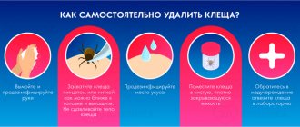
Once the insect has been successfully removed from the body, it is important to follow these steps:
- To treat the wound, it is necessary to use a soap solution, medical alcohol, iodine or other antiseptic.
- Place the tick in a container, close the lid and send it to the laboratory to identify the infection.
- The affected person must take a blood test to determine antibodies to those infections carried by this parasite. You should donate blood no later than 10 days from the moment of the bite.
- Remember the date of the bite and monitor for possible symptoms for a month. If during this period there is itching, redness and an increase in body temperature, you should visit an infectious disease doctor.
How to suspect a tick in a dog
The danger of dog ticks lies in the blurred symptoms. Signs of parasite damage to your pet may appear several days, even weeks after the first bite. During this period, the parasite significantly harms the animal and is capable of infecting various ailments.
In addition to the fact that a dog can pick up a tick, active animals often swallow ticks while playing or cleaning their fur. It is necessary to treat a dog not only if there are obvious deviations in its condition, but also if there is the slightest sign of something wrong:
- increased body temperature (more than 40 degrees);
- inhibited reactions, lack of former activity;
- weight loss, lethargy unusual for dogs;
- poor appetite, refusal of treats that previously caused delight;
- mucous membranes become pale and take on an unhealthy tint;
- the animal has constant thirst;
- the presence of convulsions, weakness in the limbs;
- bleeding in the eyes.
These and other signs are an alarming signal to the owner that the animal has a problem. In advanced cases, it is recommended to immediately take your pet to a doctor; self-help can harm the dog. Some tick-borne diseases are also dangerous for humans; the risk group includes small children and the elderly.
Signs of infection with dangerous infections
| Infection | Main features |
| Tick-borne encephalitis |
|
| Tick-borne borreliosis |
|
| Hemorrhagic fever |
|
Addresses of laboratories where you can submit a tick for analysis and have a person tested
In almost every city there are organizations and institutions (public, private) where you can take the pest for research.
Moscow
If you manage to remove a tick at home from a person, you can contact the Center for Hygiene and Epidemiology. It is located at the address: Grafsky lane, building 4, building 2. Phone number for more information: 8-495-687-40-47.
MoscowGrafsky Lane, 4k2 — Yandex.Maps
Mytishchi
Here they accept ticks that they had to remove themselves: Semashko street, building 2. Opening hours: from 9 to 16 daily on weekdays. The organization carries out all research. On weekends, ticks are also taken, but no other manipulations are carried out.
MoscowSemashko Street, 2 — Yandex.Maps
Vologda
Address of the FBUZ "Center for Hygiene and Epidemiology": st. Yashina, 1a.
Center for Hygiene and Epidemiology in the Vologda RegionSanitary and Epidemiological Service in VologdaMinistries, departments, public services in Vologda
Vladimir
If you had to tear off a parasite, you need to take it to the address: Center for Hygiene and Epidemiology, st. Ofitserskaya, 20. Phone number: (4922) 54-16-38.
Federal Budgetary Health Institution Center for Hygiene and Epidemiology in the Vladimir RegionSanitary and Epidemiological Service in Vladimir
Railway
You will have to contact the neighboring city of the Moscow region - Mytishchi. There are no laboratories for receiving ticks directly in the city of Zheleznodorozhny yet.
Izhevsk
The parasite, which had to be removed from the skin, must be taken to the regional hospital: st. Labor 17. Phone number: 21-92-24.
Republican Clinical Infectious Diseases HospitalSpecialized Hospital in IzhevskHospital for Adults in Izhevsk
Krasnodar
The Center for Hygiene and Epidemiology is located at: st. Gogol/Rashpilevskaya, 56/1/ /61/1. Telephone.
Center for Hygiene and Epidemiology Sanitary and Epidemiological Service in Krasnodar
Novosibirsk
GBUZ NSO "DGP No. 1" (220 Krasny Ave.), room 211. Phone numbers: 216-47-42, 228-71-90.
GBUZ NSO Kkdp No. 27 Children's clinic in Novosibirsk
Permian
If you had to remove the parasite, you need to contact the street. Lebedeva, 26. Contact phone.
Laboratory of Natural Focal Particularly Dangerous and Viral InfectionsMedical laboratory in Perm
Address: st. 50 years of the Komsomol, building 16.
HemotestMedical laboratory in PodolskDiagnostic center in Podolsk
Saint Petersburg
Address: Kondratievsky pr., 62, bldg. 6. Phone number: +7 967 342-18-09.
LabtestMedical laboratory in St. PetersburgMedical center, clinic in St. Petersburg
Minsk
Address: st. Filimonova, 23. Phone number 268-04-41. Parasite research is carried out for a fee.
Republican Scientific and Practical Center for Epidemiology and MicrobiologyResearch Institute in MinskMedical center, clinic in Minsk
Acariform mites
True acariform mites are distinguished by their small body size (from 0.1 to 2 mm). Their mouthparts are of a gnawing or piercing type. There is no circulatory system. These mites go through all stages of development: eggs, larvae, three nymphs and adults. Many acariform mites are the causative agents of scabies in domestic animals. Some of them are involved in the development of ruminant anoplocephalids as intermediate hosts.
Depending on the belonging of scabies pathogens to different genera, it is recommended to establish a diagnosis not for scabies as a whole, but for acarosis, psoroptosis, chorioptosis and notoedrosis.
These photos show what cariform mites look like:
Caring for your dog after tick removal
If you manage to rid your dog of ticks yourself, you need to:
- Inspect the wound to see if the arthropod's head remains inside. If so, the area needs to be disinfected and the residue removed with tweezers. If this is not done, there is a risk that an inflammatory process will begin. In this case, it is inconvenient to work with a needle; you can pick the wound too much.
- If the head cannot be removed, the wound is regularly lubricated with iodine. The dog’s body itself will reject the foreign body after some time.
- If the wound is clean, it is treated with any antiseptic: brilliant green, iodine, peroxide, alcohol.
After the parasite is removed, there is no point in immediately taking the dog to the veterinary clinic and removing the tick from the dog for testing. On the first day, protozoa are not detected in the peripheral blood. Therefore, the result of the analysis will be negative, but also false. Accurate results appear within 3-4 days. And even with a negative indicator, a repeat analysis is done on days 5-7.
It is also impossible to determine from the ectoparasite itself whether it is a carrier or not. An infected tick does not look different from a normal tick. Even if he is a carrier, it is not a fact that he was the one who managed to infect the dog.
Females drink the blood of animals. Having sucked, they do this for 5-7 days, inflate and become like a large dark pearl. Then the female falls off her body - she needs to lay eggs in a secluded place to continue the race. Therefore, if a small, newly attached tick is found and the dog develops symptoms of the disease, then the culprit may not be this one, but a completely different specimen that has fallen off long ago. After all, symptoms of infection can appear from 3 to 21 days after the bite. The owner only has to monitor the pet and measure the temperature up to 5 times a day.
What happens if you don't remove the leftovers?
Even if you remove the tick very carefully, it may happen that part of it comes off and remains in the skin. It is highly undesirable to leave it there. The head of the parasite is a non-sterile foreign body. Its presence in soft tissues can provoke the development of an inflammatory process and suppuration. In rare cases, symptoms of intoxication appear: nausea, weakness, dizziness.
To avoid this, you should try to remove the remains of the parasite, and if you cannot do this yourself, seek medical help.
What to do if the tick's head remains in the body of a cat or dog
Before going to the veterinarian, you should try to remove the insect's head. The needle should not be used on animals: the pet may become frightened and jerk sharply, which will lead to the expansion of the wound and the entry of bacteria into it. Only tweezers can be used.
If the head is stuck deeply and does not stick out above the skin at all, the tubercle formed at the site of the bite is treated with iodine. This is where home actions after a tick bite end.
The veterinarian will cut the pet's skin under local anesthesia and remove the head of the tick. It is advisable to send the insect for analysis. The cat will be given an injection to boost immunity, and, if necessary (if the tick turns out to be a carrier of the disease), other therapeutic injections will be given. Therefore, it is better to stay with your pet in the city for several days after the bite.
If you do not treat the tubercle with the remaining tick head in it, inflammation will develop within 1–2 days. It is accompanied by suppuration of the wound, the appearance of redness, and occasionally, hair loss in the area of the bite. The animal may develop intoxication if the area of inflammation becomes extensive, as well as fever.
Another danger for a pet is viral diseases that are transmitted through the salivary glands of a tick. They can lead to the development of a crisis within 12–24 hours from the moment of the bite, which will end in death.
Therefore, immediately after discovering a tick, you should go to the veterinarian. Some viral pathologies do not lead to death within a few days, but in an advanced state they can also lead to death.
It is important not only for city dwellers, but also for rural residents to know what a drunk tick looks like and what to do if this small insect bites you. There are a large number of ticks in nature, but not all of them pose a threat to human life and health
The most dangerous of them is. It is the source of such terrible diseases as encephalitis, borreliosis or hemorrhagic fever. Costs .
This insect constantly migrates from one place to another, so it is not always possible to say with certainty which species live on your territory. Traditionally, the tick lives in forests and where trees grow densely. A garden, square or just planting can become a source of habitat. There are a lot of ticks in places where it is dark and humid. They sit in the grass, on the leaves of trees and bushes. There are especially many of these individuals in deciduous and mixed forests.
These little pests love paths, garden paths, and roadsides where there is a lot of dried grass. You should be especially careful when walking on forest edges, in ravines or near forest streams. There is a high probability that you will be bitten by an insect in a willow thicket, in a birch grove, in the grass near a river. In places where there are large crowds of people, you can easily find a tick. As scientists have proven, bloodsuckers are attracted to the natural smell of a person or animal, and their sensitivity is very developed. The tick recognizes odors at a distance of more than 12 m.
How to properly remove a tick from your body
What happens if you tear off a tick depends on how professionally the procedure is carried out. It is necessary to remove the parasite carefully and strictly according to a certain algorithm. Otherwise, the following complications may occur:
- the head or proboscis of the tick is stuck in the skin;
- the parasite will be squeezed, which will lead to the spread of the poison;
- the insect will suffocate or otherwise die, increasing the risk of inflammation and infection.
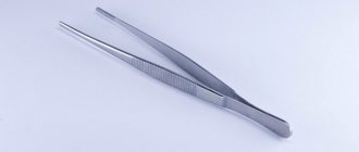
To avoid these problems, you should use one of three methods to remove the parasite. It is important to understand that the best way to eliminate a tick is to use special tools. They can be purchased at the pharmacy.
A number of instruments (working like tweezers) are equipped with test tubes into which the parasite can be placed to be tested. This is required if a person lives or vacations in an area where the encephalitis tick is spread.
- The first method of removal is twisting. It is necessary to squeeze the head of the tick with your index finger and thumb. In this case, under no circumstances should you put pressure on the body itself. When the fingers are securely fixed enough, you can twist the parasite. This can be done clockwise or counterclockwise. The main thing is that the parasite is located perpendicular to the skin, otherwise the proboscis may break off. This method is the most convenient and fastest. Experts advise using tweezers rather than fingers, if you have them at hand. And the best option, as already mentioned, is to use a special tool. It is advisable to avoid skin contact with the tick by wrapping your fingers with a piece of cloth soaked in alcohol.
- The second method is removal with thread. It should be strong and not tear from being pulled. The thread is wrapped around the tick near the wound. You can't grab the abdomen. After application, the thread is tightened and light tugging movements begin. This method is not suitable for removing insects from pets, as it may take several minutes. It also does not always work on small mites.
- The last method, the most controversial, is the use of compounds with a high percentage of fat content, designed to suffocate the insect. You can use olive or vegetable oil, or any similar liquid. It is carefully applied to the wound so that the substance gets inside and affects the head of the tick. The danger of the method is that the parasite can actually suffocate, and not just feel discomfort and crawl out. In this case, the dead insect will become firmly stuck in the skin, inject poison into the blood and lead to the development of inflammation.
The correct algorithm for removing a tick from a dog
To timely detect a parasite on a pet’s body, it is important to regularly examine the animal after each walk during the peak of parasite activity. For these purposes, you must have a comb, which must be used to comb the coat against hair growth.
Particular attention should be paid to places hidden from view - the neck, ears, groin and elbow areas. Ixodid ticks also like to localize on the head and back
If the tick has already burrowed into your pet’s body, you can contact a veterinary clinic or try to properly remove the tick from your dog at home. To do this, you must follow a number of recommendations:
- First of all, you need to protect yourself by wearing gloves. The tick can be infected and infect a person with Lyme disease (borreliosis) or encephalitis.
- Do not try to remove the parasite from the dog's body. The oral apparatus of a piercing-sucking type tick is equipped not only with a proboscis, but also with a whole set of spines. When trying to tear off a tick, the head of the parasite may remain inside and provoke the development of an infection or inflammatory process.
- Try not to panic and calm your pet. It is necessary that the room has good lighting. This will allow you to carefully examine whether, after removing the tick, the head of the parasite remains in the epidermis.
Pincers
Specialized animal stores sell special tick pullers that greatly simplify the process of pulling out ticks. They work on the principle of a nail puller. The tool is brought close under the protruding body of the parasite and begins to rotate until the tick comes out completely, along with the head.
Threads
Not everyone knows how to use a thread to pull out ticks. There is a high risk of cutting the thread from the head of the tick to the body, thereby leaving dangerous fragments of the parasite under the skin. Many owners make the mistake of using a thread when pulling out a tick. A regular thread can cause more damage than tweezers. In order to correctly carry out the procedure for removing a tick, you need to carefully make a loop out of a thread, carefully place it over the body of the tick and tighten it slightly, but not completely. Next, you need to make loosening movements, slightly tightening the thread. Within 10-15 minutes the tick will begin to leave its shelter.
Insulin syringe
In order to remove a tick that has attached itself to your pet, you can use an insulin syringe. First remove the needle from the injector, lubricate the tip with oil and apply it to the skin so that the entire body of the parasite is in the syringe. They begin to gently pull back the piston and, under the influence of a vacuum, the tick will leave the skin. After the owner has successfully pulled out the tick, there is no need to relax. The dog should be closely monitored for at least 3 days after the bite. If symptoms such as high temperature, febrile conditions, change in the color of urine, refusal to feed, icterus (yellowness) of visible mucous membranes appear, you should contact a veterinarian for help as soon as possible.
Tweezers
When squeezing a tick with tweezers, there is a high probability that more infectious agents will enter the dog's bloodstream. Therefore, this method is not recommended. tweezers.
Vegetable oils and gasoline
Experienced dog breeders and veterinary specialists do not recommend using vegetable oils or gasoline to remove ixodid ticks from a dog’s body.
Ticks of the family ixodidae (Murrey, 1877)
Morphology of ixodid ticks. More than 49 species of ixodid ticks have been registered in the CIS. All of them are temporary ectoparasites of vertebrate animals and carriers of pathogens of many diseases of mammals and birds: arboviruses, bacteria, blood parasites, rickettsia, etc.
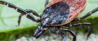
Ixodid ticks are the largest; the size of females that have drunk the blood of animals can reach 20-30 mm. Segmentation of the body is not pronounced; the head, thorax and abdomen are fused into a continuous, undivided body, resembling a flattened sac, narrowed anteriorly. In the front part of the body there is a proboscis - the gnathosoma, followed by the idiosoma (the body itself). The proboscis, an organ of fixation, consists of a base, or collar, a dense chitinous plate of three-, four-, five- or hexagonal shape; pairs of upper jaws (chelicerae), which serve to cut the skin of the animal; hypostome (fused lower jaws), shaped like a harpoon, covered with teeth directed backwards for strong attachment to the host’s body; two four-segmented palps (palps), acting as organs of touch. At the base of the proboscis, females have a special sensitive organ - pore fields.
Adult ixodid ticks exhibit pronounced sexual dimorphism. On the dorsal side of the tick idiosome there is a scutum (scutum) - a thick, shiny chitinous formation. In males, the scutellum covers the entire dorsal side, while in females it covers only 1/3 of the idiosome behind the proboscis. This feature is the main difference between females and males. The color of the shield is often dark brown or gray. There are grooves on the scutum, the color, shape and number of which have diagnostic significance. The body color of drunken females varies greatly from steel-blue to light gray with a brownish or pinkish tint. The color of ticks that sucked lymph is always lighter than those that fed on blood.
In ticks of the genera Boophilus, Dermacentor, Rhipicephalus, Hyalomma, eyes are located on the sides of the shield, approximately at the level of the second pair of legs.
On the ventral side of the idiosome, the mite has a genital opening (at the level of the first–second coxa) and an anus (far behind the fourth pair of legs). In all genera (with the exception of Boophilus), an anal groove runs near the anus, going around it in front (only in Ixodes) or behind (in all other genera). Four pairs of jointed legs are attached to the idiosoma on the ventral side. Adults and nymphs have four, and larvae have three pairs of legs, consisting of five segments: a coxa (coxa), fixedly attached to the body, a trochanter, a thigh, a tibia, and a tarsus, which has a sucker at the end and two claws. The body (together with the scutum) and limbs are covered with a varying number of bristles, which, together with suction cups and claws, serve the mites for crawling and attaching to the animal’s cover and human clothing; The bristles serve as organs of touch. On the paws of 1 pair of legs there is an organ of smell - Haller.
The internal structure of ticks includes the digestive, respiratory, excretory, reproductive and nervous systems.
The digestive apparatus begins at the base of the hypostome with the oral opening, followed by the pharynx, which gradually passes into the esophagus, which flows into the central intestinal canal with long convoluted blind branches extending from it. The small intestine extends from the midgut, connecting, like the hind intestine, to the rectal bladder, where excretory products accumulate. The rectum opens with the anus, closed by a special valve. One female can suck 2-3 ml and stock up on a large amount of blood necessary for the formation of eggs. On the sides in the front of the body there are paired cluster-shaped salivary glands; The saliva produced during feeding is excreted through the salivary ducts through the mouth into the host's wound. Saliva has lysing properties, prevents the clotting of blood entering the intestines, and anesthetizes the bite.
The excretory apparatus consists of one pair of long, thin tubes (Malpighian vessels) winding throughout the body cavity. One end of them is blind, lies freely in the body cavity, the other flows into the rectal bladder.
The respiratory apparatus consists of a dense network of respiratory tubes - tracheas, extending from the common and cephalic trunks. A dense network of branched tracheas covers all parts of the body and opens outward along the edges of the body behind the fourth pair of legs with two spiracles (stigmas) located on special plates - peritremes (their shape is taken into account when identifying ticks).
The circulatory system is not closed. The blood of ticks (hemolymph) is colorless, washes all the organs of the body and enters the heart, which has the shape of a thickened sac. The aorta and trunks extend from the heart, supplying blood to the legs, proboscis and brain.
The nervous system is a single solid mass located in the front of the body on the ventral side. Many nerves extend from the brain to the limbs, eyes, mouth and internal organs.
The sense organs include the eyes (with the exception of the genus Ixodes and Haemaphysalis), a special organ of smell - Haller's organ, located on the paws of the first pair of limbs, as well as tactile bristles covering the body, proboscis, and limbs.
The female reproductive organs consist of the ovary, oviducts, uterus, spermatic receptacle, vagina, and a pair of accessory glands. In addition, there is a Zhene organ that produces a secretion that lubricates the eggs during laying. In males, the genital organs are represented by the testes, vas deferens and accessory glands.
The development cycle of ticks goes through transformations (metamorphoses): egg, larva, nymph and adult – sexually mature females and males. The female lays from 3-4 to 10 or more thousand eggs and dies (one gonotrophic cycle). After a certain period of time (1-2 weeks), depending on temperature and air humidity, the eggs hatch into six-legged larvae measuring 1.0 × 0.5 mm. Some time after the larvae emerge from the eggs, they become ready to attack animals and suck blood. The drunken larva increases in size and after 5-10 days molts into a nymph with four pairs of legs. The size of a hungry nymph is 2-3 mm. For subsequent development, the nymph must also drink blood, and then it turns into the adult phase - male or female. Ixodid ticks feed on the host's body a certain number of times over several days in each active phase. Depending on the type of food, one-, two- and three-host species are distinguished.
In single-host ticks (Boophilus calcaratus, Hyalomma scupense), the larva, nymph and mature individuals feed on the same animal on which they molt. Fertilization of females by males occurs in animals during the sucking period. Only well-fed, sexually mature ticks leave their host. The fallen females look for suitable places in crevices, under stones, under dead parts and roots of plants, under dry manure, etc., lay eggs and die.
In two-host ticks (some species of the genera Rhipicephalus, Hyalomma), development occurs on two hosts. Larvae and nymphs feed on the former. Here the larvae, without detaching, turn into nymphs on the spot, which, after feeding, fall off the host to the ground and turn into mature ticks. The latter attack the animal - the second owner, having fed, leave it to lay eggs on the ground.
In three-host ticks (genus Ixodes, Haemophysalis, Dermacentor, some species of the genera Hyalomma and Rhipicephalus), all three phases - larva, nymph and mature individuals - feed on three different animals, i.e., each phase leaves the host’s body after the end of blood-sucking. Larvae and nymphs feed on the blood of small mammals.
The lifespan of ticks varies. Larvae are capable of fasting from several months to a year, sexually mature individuals of some species - for several years. Hungry adults, drunken nymphs, and tick eggs overwinter in nature.
METHODS FOR COLLECTION AND STUDY OF IXODID TICKS
Methods for collecting ticks are divided into epizootic and regular, universal and selective, complete and partial. Epizootic collections are usually used to determine the degree of well-being of an area (farm) in relation to ticks; they are carried out both in natural and guest biotopes. Regular census collections and observations are carried out either continuously for a certain period of time, or at intervals of several minutes and hours, 5-10 days, and even up to several months. Collections can be general in case of a small number of animals and selective in case of a large number of animals. A harvest is called complete when all visible parasites are caught on animals. During partial collection, only an arbitrary number of parasites are counted and counted on a conventional scale designated: “very many”, “many”, indicating approximately the number for each designation.
The direct collection method involves partial or complete collection of ticks found on animals. When collecting from animals with short and straight hair, use a thin wooden plank (size 15-20 × 5 cm) with pointed long edges. It is carried out against the hair, looking at the skin behind the plate for the presence of ectoparasites.
A survey of the area for the presence of ticks is carried out in order to identify inhabited and uninhabited areas, the boundaries of these areas, species composition, the dynamics of the seasonal number of these arthropods in pastures, etc. (for this purpose, devices are used: a drag net, a flag, a towel, a screen, etc.) . The area can be examined using the method of route epizootic and regular tick counts in open nature, near livestock buildings, runs, watering places and resting places for animals. All received data is plotted in the form of symbols on a schematic map of the farm territory.
The best preservative liquid for ticks is 70% alcohol, 4-5% aqueous solution of formaldehyde. All ticks isolated from each count (from an animal, from pastures, etc.) are placed in a separate container, and the appropriate label is also placed there. During further processing, ticks are divided into genera and species, and then the number is calculated for 10 days or more. For greater clarity, the results obtained are depicted in the form of curves and diagrams.
When collecting ticks, you should reliably protect yourself from attacks by parasites. To protect your hands, you should use mesh handcuffs impregnated with a composition containing phthalate. The sleeves are made from a piece of mesh measuring 35x25 cm, trimming the edges with braid and sewing them in the form of a cylinder with a diameter of 18 cm. The sleeves are tied to the cuff of a tunic, shirt, etc.
DIFFERENTIAL DIAGNOSTICS OF TICKS OF THE FAMILY IXODIDAE
1. GENUS BOOPHILUS , Curtice, 1891. Body length of hungry individuals is 15 mm. The color of the scutellum is from yellow-brown to light brown. The eyes are flat and lateral. The proboscis is short, its base is hexagonal. The palps are short and ribbed. Males have two pairs of scutes on the ventral side of the idiosome. In the CIS it is recorded in the forest-steppe of southern Ukraine, the North Caucasus, Kazakhstan, Central Asia and Transcaucasia. Biotopes include river valleys, damp meadow thickets, and livestock buildings. The feeders are large and small cattle and horses. Active period from spring to December.
Boophilus calcaratus, Birula, 1895 – single-host tick. It is a carrier of Piroplasma bigeminum, P. ovis, Babesia colchica, brucellosis, Q fever.
2. GENUS HYALOMMA , Koch, 1844. Ticks are the largest in the family. IXODIDAE. The proboscis is long, its base is rectangular, the coxae of the first pair of legs are deeply split. Males have 2-4 pairs of scutes on the ventral side. The peritrema is occipital or retrovidous. The legs are powerful and long. Their biotopes are steppe, desert and semi-desert landscapes; some species live in shrubs, open forests, lowland and mountain forests, and in livestock buildings. They are carriers of pathogens of piroplasmosis, possibly tick-borne typhus, hemorrhagic fever, human Q fever, plague, leptospirosis and animal brucellosis. From this genus, farm animals are parasitized by the species H. anatolicum, H. detritum, H. plumbeum, and H. scupense.
H. Anatolicum, Koch, 1844, Found in floodplain meadows, fallow lands, livestock buildings near the nests of synanthropic birds in Central Asia, Kazakhstan, Transcaucasia and the Northern Caucasus. The hosts are cattle, horses, sheep, etc. The tick is a three-host tick. Adults are found on animals in April-May-June; larvae - in July-August, nymphs - in autumn and winter; parasites have one generation per year. The tick is a carrier of Theileria annulata, Th. mutans.
H. Detritum, Schulze, 1919, carrier of Theileria annulata, Th. mutans. The northern border of its range runs through Transcaucasia, Southern Kazakhstan, and throughout Central Asia. The tick has two hosts: larvae and nymphs parasitize one host, and adults parasitize another host. Active in May-September, all agricultural animals can be feeders. One generation develops per year.
H. Plumbeum, Koch, 1844, is found in intermountain valleys, deciduous forests, forb fallows, stacks and haystacks in the south of Ukraine, in Transcaucasia and Central Asia, in the south of Kazakhstan. The hosts are large and small domestic and wild mammals (imago) and brown hare, wild and domestic birds, mouse-like rodents (larva and nymph). The tick has two hosts and feeds on animals from July to October. It is carried by Piroplasma caballi, Nuttallia equi.
H. Scupense, Schulze, 1918, is a single-host tick. The northern border of the range passes in Ukraine, in the Kursk and Saratov regions, the Lower Volga region, and Central Kazakhstan. To the south, ticks are found everywhere in mixed-grass floodplain meadows. They parasitize animals during the cold season (October-May). Vector by Theileria annulata, Nuttallia equi.
3. GENUS DERMACENTOR , Koch, 1844. Characteristic of all species is the presence of silver-white (light enamel) spots on a dark background of the dorsal shield (marble pattern), limbs and proboscis. The proboscis is short, the palps protrude beyond the rectangular base of the proboscis. The eyes are flat and marginal. Males without ventral scutes. All species develop according to the three-host type. Ticks have one generation per year, the imago lives up to two years. They live in forests, steppes, and semi-deserts; in mountainous areas at an altitude of more than 2000 m above sea level they are carriers of pathogens of piroplasmosis, tick-borne encephalitis, tick-borne rickettsiosis, plague and tularemia.
D. marginatus, Sulzer, 1776 – carrier of Piroplasma caballi, Nuttallia equi, A. marginale, A. ovis, Th. recondita. Ticks are found in Ukraine, Moldova, Crimea, Voronezh region, the Middle and Lower Volga region, Kazakhstan, the North Caucasus, the southern part of Western and Eastern Siberia and Central Asia. Larvae and nymphs feed on mouse-like rodents, hedgehogs, water rats and other small animals. The adult parasitizes cattle, sheep, goats, and horses. The tick attack begins in May (mainly by adults); in the summer, mature ticks are not found; in the fall, the greatest increase in the number of ticks in animals is observed.
D. Pictus, Hermann, 1804 – carries P. caballi, N. equi, P. canis, A. marginale, equine encephalomyelitis virus, Omsk hemorrhagic fever virus. The distribution area is from the western borders to Eastern Siberia. Its biotopes are confined to the forest zone and meadow forest formations. The adult attacks animals at the end of March – April – May. Larvae and nymphs feed on hedgehogs, hares, and mouse-like rodents.
4. GENUS RHIPICEPHALUS , Koch, 1844 – relatively small ticks (hungry 2-5 mm, drunk females 10-15 mm), reddish-brown in color. The eyes are flat, marginal, and inconspicuous. The proboscis is short in the form of a truncated cone, its base is hexagonal. The palps are short, the coxae of the first pair of legs are deeply split, the anal groove goes around the anus from behind. Males have two pairs of ventral scutes.
Rh. Bursa, Canestrini Et Fanzago, 1877 – carrier of B. ovis, P. ovis, A. ovis, Th. recondite, A. marginale. Distribution area: Crimea, Krasnodar Territory, North Ossetia, Transcaucasia, Turkmenistan. Tick biotopes include virgin pastures and pastures with sparse shrubs. The tick has two hosts. In the adult phase, it parasitizes cattle and small cattle in March-July, the larvae attack in the second half of summer and autumn, the nymph parasitizes animals during winter. There is one generation per year.
5. GENUS HAEMAPHYSALIS , Koch, 1844 - mites with a short proboscis having a quadrangular base; palps are wide. The dorsal shield is brownish-brown or dark brown without a marbled pattern. There are no eyes. The anal groove goes around the anus in a closed semicircle from behind. Males without anal scutes; females without lateral grooves. Three-host mites. They transmit pathogens of piroplasmosis, brucellosis, tularemia and a number of rickettsial and viral diseases of animals, human plague. The following species are of greatest veterinary importance:
H. Otophila, Schulze, 1915 – carrier of A. ovis, P. ovis. It lives mainly in the arid steppes of the plains and foothills of Ukraine, the Krasnodar and Stavropol territories, Chechnya, Ingushetia, Dagestan, Transcaucasia, and Turkmenistan. The adult parasitizes small cattle and cattle in October-March. Larvae and nymphs feed on hedgehogs, hares, and steppe birds.
H. Japonica, Nuttall et Warburton, 1915 – carrier of Th. sergenti. It lives in the Far East in coniferous-deciduous forests. The concentration of ticks increases along river valleys and at the junctions of landscapes. The adult parasitizes animals in the first half of summer; larvae and nymphs feed on cattle, sika deer, etc. All phases of development attack animals.
6. GENUS IXODES , Latreille, 1795 – long-proboscis mites. The base of the proboscis is quadrangular in shape. The chitinous cover of the female is of various shades - from gray to red-pink-brown. There are no eyes or scallops. The anal groove (unlike other genera) goes around the anus in front in a closed semicircle. The first pair of cokes is not split. All species develop according to the three-host type.
I. Ricinus, Linnaeus, 1758 and I. Persulcatus, Schulze, 1930 are carriers of B. ovis, B. caucasica, A. ovis, A. marginale, pathogens of human viral and bacterial infections. Distributed in the European part of our country, with the exception of treeless steppe zones in the south, Murmansk and Arkhangelsk regions in the north, and the species' range in the east is limited by the Volga River. In the foothills and mountains of the Caucasus, ticks are often found in areas dominated by forests and shrubs.
Adults parasitize cattle, small cattle, horses, wild mammals and birds. Larvae and nymphs feed on all types of small mammals and birds. The life cycle of ticks in the north takes 3-4 years, in the North Caucasus - within one year.
THE FIGHT OF IXODID TICKS is carried out by destroying them in biotopes and on animals.
Destruction of ticks in biotopes is the most effective method. In most species of ticks, egg laying, larval hatching, and molting developmental phases occur on the ground (in natural pastures). Therefore, plowing and creating pastures with seeded grass can dramatically reduce the number of ticks, while the composition of vegetation, temperature and soil moisture change, which prevents the development of ticks. Sometimes changing pastures gives satisfactory results.
An important place in the fight against ixodid ticks is their destruction in biotopes - in livestock buildings by sealing cracks, rodent burrows, and destroying vegetation around farms. Then the room is treated with a 1% suspension or emulsion of sevin or dicresyl at a rate of 200-400 ml/m2. Before placing the animals (3-4 hours before), the room is ventilated, then the feeders are washed with hot water and the remaining drugs are removed. If ticks are found on animals, they are treated with acaricides.
Animal processing methods. Six main methods are used: bathing in floating baths, spraying, dusting, watering along the spine, applying acaricides to the coat in the form of a paste, injections and oral use of acaricides. The first two methods are acceptable only in the warm season, the rest - in both warm and cold periods. Sheep are usually bathed (see tables 1 and 2) .
Spraying is carried out on specially equipped sites - MDU, with automatic supply of acaricides, or on fenced concrete sites, using spray equipment: LSD, DUK, VDM, etc. Working fluid is used for small livestock - 1.5 liters, for cattle and horses – 2-4l, for camels – 5-10l per animal. Acaricides contained in aerosol and propellant cans are also used for spraying. The treatment is carried out from a distance of 20-30 cm, pressing on the head of the aerosol can or actively working with the lever of the propellant can.
Preparations for irrigation - purons are applied to the skin of the animal along the spine; When processing sheep, the fleece is first spread apart. In regions where vaccination or mitigating chemoprophylaxis is used, animals are treated once every 15-20 days, and with pencils or Butox - after 25-40 days, depending on the residual effect of the drug. In the cool season, it is advisable to use dust, pencils, aerosols, and acaricide injections.
Animals are treated with acaricides before ixodid ticks attack them, for which they use drugs that act simultaneously on larvae, nymphs and adults. The effectiveness of the drugs is determined by the absence of newly attached larvae, nymphs, and adults by the time of the next treatment. Acaricides can cause toxicosis in animals, so anti-tick treatments are not recommended for young animals up to 3 months of age and for pregnant animals one month before giving birth. To avoid toxicosis, animals are processed in the cool part of the day.
When using acaricides, it is very important to calculate the dose and concentration of the drug according to the active substance (AI) in working emulsions and solutions. When calculating, use the following formula:
X=AxB
C
Where X is the amount of technical preparation (emulsifying concentrate) required to prepare the working solution, kg; A is the amount of solution that needs to be prepared for processing, l; B is the concentration of acaricide according to the DV, which must be obtained in the working solution; C-content of DV in the original preparation in %.
Calculation example: from 20% emulsifying concentrate (e.c.) propoxur for spraying 250 heads of cattle, it is necessary to prepare 0.5 x 250 = 125 l of emulsion of 0.4% concentration. Using the formula we get: X=(125x0.4)/20=2.5. Therefore, to prepare 125 liters of 0.4% propoxur emulsion, you need to take 2.5 kg of 20% e. to this drug.
In addition, traps have been developed to catch imagoes and nymphs of ixodid ticks on the host’s body. The trap is disc-shaped and contains ferromone and acaricide. The trap is attached to the tail. Acaricidal collars and bags with the acaricide amitraz or cyhalothrin, which are attached to the tail of the animal, are also proposed. The effectiveness of the bags reaches 87-93% and remains at this level for 90-94 days.
Ways to remove ticks at home
They are considering different options, the choice depends on the conditions under which the pest has to be pulled out.
Tick remover - any available one purchased at a pharmacy
A short algorithm that allows you to get arthropods in different conditions:
- Install the tool so that the parasite is located between the protruding elements.
- Make screw movements (turn the twister several times).
- To get the tick, you do not need to make significant efforts, as it can come off. It is quite easy to pull the parasite towards you. It must be taken into account that he will not crawl out on his own.
Video: what a twister looks like
Reaching with tweezers
The principle of removing the parasite is the same as in the previously discussed case, but the method of capturing the pest is different
So, you need to be careful when unhooking an arthropod using tweezers. To remove a tick from a person at home, you need to have experience using such a tool.
Video: how to remove a tick with tweezers|How to quickly and safely remove a tick from a human body!
Removing a tick with your fingers
The procedure is performed with gloves, for this case this is a mandatory measure. The fingers are wrapped in gauze, which will reduce the intensity of the pest’s sliding on the latex. To remove a tick from a person, you need to make screw movements. You should first grab the parasite with your fingers, but you cannot squeeze it, otherwise the head may break off.
Video: how to remove a tick with one finger!
Pulling a loop from a thread
You need to tie the thread so that you can gradually tighten the loop around the tick (a knot is not recommended). They throw a thread over the parasite, fix it, and begin to twist it. To do this, you need to pull the opposite ends in different directions. Moreover, this way you can damage the parasite with a thread.
Video: how to pull out a tick with a thread if bitten by a tick
How to remove a tick with a syringe
Prepare an insulin syringe without using a needle. Cut off the lower part (the narrowest part), but do not touch the piston inside. To properly remove a tick at home, you need to place the syringe exactly above the arthropod and press firmly. Then the piston is raised. As a result, the tick will gradually emerge from the epidermis.
Video: Is it possible to pull out a tick with a syringe - experiment
Using oil
In this case, it is enough to apply a small amount of the product to the tick and the bite site. The oil will block the pest’s respiratory tract, and it will try to get out on its own. In this case, the tick can release saliva in large volumes. As a result, the risk of infection will increase.
Video: how to pull it out with oil - experiment
Trix Tix Lasso Loop
It is safer to use this device. You need to press the button located on one side. When the tick is fixed, it is released, which ensures fixation of the parasite. Extraction is carried out by turning the plier in one direction, the action is repeated several times. Then you need to pull the pest towards you. This should be done so that no fragment of the parasite’s body remains in the epidermis. Removing it is more difficult than removing an entire pest.
Video: removing ticks using a loop
Pro Tick Plate
This is a compact tool, the mechanism of action is no different from a twister. But these devices have different designs. The plate is straight, on one side there is a slot for fixing the pliers, on the other there is a hole for attaching to a keychain to be worn with keys.
Video: how to remove a small tick from a person with a special tool
How to get rid of a tick with a bank card
A sharp knife must be used. A proper plastic card extractor should have a slot, it is cut in the shape of the letter V. You can get such a device for free, and its effectiveness can be compared with other tools. If a dog or taiga tick has bitten, it is fixed using a card (the parasite must be in the slot). Then they begin to twist it out, after which they remove it; to do this, you need to pull the parasite towards you.
Video: how to remove a tick with a plastic card
- you need to touch the tick with a lit cigarette;
- A needle is inserted into the body of the parasite, which leads to its death, after which it is easier to remove the pest.
Tick extractor
To remove ticks, use both special tools and improvised means. Improvised means such as tweezers, thread or a syringe will not guarantee safe removal of the tick, and moreover, they can only aggravate the situation. Also, never try to pull out a tick with your bare hands - the tick can be easily crushed, and then viral particles can easily penetrate into the wound. Use KleschKart effectively and safely:
- KleschKart is a plastic card with a special groove. The groove must be placed under the body of the attached tick, without pressure or effort, move the card forward and remove the parasite; as a result of this manipulation, you will get it alive and intact.
- The kit includes an antiseptic wipe for treating the bite site and a test tube for the tick (to deliver the parasite to the laboratory for analysis). The KleshKart easily fits into a wallet, business card holder or pocket.
During tick season, we are all at risk. a Tick Card with you , inspect your body after walks. Take care of yourself!
How to pull out the proboscis
Not only the whole head of the tick, but also its proboscis may remain under the skin. Take a closer look - if a black dot is visible at the site of the bite, then this is most likely the proboscis. If there are no signs of suppuration, you can get rid of the parasite's proboscis. Just pull it out like a regular splinter - with a needle or pin.
- Clean the sharp needle and the skin around the bite site with rubbing alcohol.
- Use a disinfected needle to pick up the sting of the tick and pull it out.
- Lubricate the wound with iodine.
If there is swelling and signs of suppuration around the bite site, then you cannot pull out the proboscis yourself - you need to consult a doctor so that he can make a correct skin incision under sterile conditions and remove the remains of the parasite.
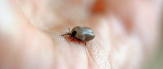
How do you know if the parasite has not been completely removed?
Once on the victim's body, the tick does not bite immediately. He can crawl from place to place for several hours, looking for the most suitable place (with thin skin and closely located vessels). Moreover, the parasite can make several targeted attempts, trying to bite through the skin.
But when the tick finally gets attached, it is not easy to pull it out. The bloodsucker does not dive deep into the skin, but firmly clings to the soft tissue with its proboscis. It is for this reason, as well as due to carelessness or clumsiness, that often during the extraction process the body of the arthropod is torn off from the head, which remains in the thickness of the skin.
Buried tick
Given this possibility, it is very important to carefully examine the wound after removing the parasite. It is advisable to use a magnifying glass. If the wound is clean, everything is fine. If dark inclusions are visible in it, then most likely this is part of the head or proboscis of a tick, which must be gotten rid of as quickly as possible.
What to do if the tick's head remains in the dog's body?
If attempts to remove the tick were unsuccessful and led to the body being torn off from the head of the insect, it is again advisable to seek help from a veterinarian, but if for some reason this cannot be done, the recommendations are as follows:
- Disinfect the bite site with alcohol or vodka.
- Lubricate the location with gasoline, kerosene, machine oil, and white alcohol. It is recommended to drip each substance drop by drop separately from each other, maintaining an interval of 5 minutes. It is necessary to ensure that the dog does not lick toxic substances!
- Then you need to lightly press down the head, and if it continues to remain in the dog’s body, it is recommended to make a loop out of thread, throw it over the insect’s head, tighten it tightly and try to pull it out. If it is not possible to pick up the head with a thread, you can use a thin, disinfected needle to pick out the parasite from the dog’s skin. When the head is completely removed, it is necessary to carefully treat the bite site with alcohol.
- After all the manipulations, you need to very carefully monitor the pet’s condition, and if alarming signs appear, such as an upset digestive system, fever, neurological disorders, etc., urgently take the dog to a veterinary clinic.
It is important to understand that not all manipulations to remove a tick or parasite remains are successful. Therefore, if possible, it is best to take the dog to the clinic
The doctor will make a small incision and quickly remove the tick or its remains - this is much safer than picking “blindly” on your own.
You need to understand that parasites can cause serious complications, and in order to protect your pet, drug therapy will be required:
- medications – Forticrab, Pirosan, Imizol;
- vitamins to improve immunity;
- heart medications - to strengthen blood vessels;
- hepatoprotectors – to restore the liver;
- in especially severe cases, blood purification from infection is indicated.
Attention! If the owner does not notice the tick or its remains, the animal will die - according to statistics, 98 out of 100 cases end in death. After removing the bloodsucker, it will be necessary to examine the animal for the presence of other ticks
In most cases, bloodsuckers attach themselves to open areas of the body - muzzle, ears, belly, neck, areas between the fingers. Therefore, these places should be inspected first.
After removing the bloodsucker, you will need to inspect the animal for the presence of other ticks. In most cases, bloodsuckers attach themselves to open areas of the body - muzzle, ears, belly, neck, areas between the fingers. Therefore, these places should be inspected first.
What not to do?
After people try to remove a tick on their own and its head comes off, many begin to panic. In such a state, you can do many wrong things that will lead to unpleasant consequences.
Signs of borreliosis in humans after a tick bite
Five simple rules to avoid making the situation worse:
- Do not lubricate the attached tick with sunflower or other oils or alcohol. He will not crawl out on his own so quickly, but will only suffocate, spitting out a huge amount of microbes along with saliva.
- Do not scratch or rub the bite area. It is necessary to endure this sensation, despite the severe itching.
- Do not apply any ointments, emulsions or creams to the affected area. Bandaging and sealing the wound is also contraindicated.
- Do not wet the bite wound for 48 hours and protect it from direct sunlight.
- Don't ignore going to the doctor. Even a previously vaccinated person against encephalitis will not save you from other dangerous infections, for example, borreliosis.
If there are doubts that there will be no problems when pulling out the parasite and its head will not come off, then it is better not to risk it and go straight to the hospital to have the tick removed. It is especially difficult to independently remove severed parts of an insect on the head, armpits, back and perineum.