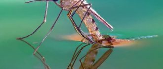
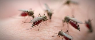
Despite the development of medicine, malaria remains one of the most common diseases in modern times. Although there are now drugs for its successful treatment, it claims the lives of almost two million people every year. Malaria is a protozoal disease and is caused by a small parasite called Plasmodium falciparum. It has a complex development cycle, and it can only exist in the body of a carrier. Therefore, you can become infected with malaria through a mosquito bite. It is the main host of Plasmodium falciparum. And man is its intermediate host.
What is Plasmodium falciparum?
The causative agent of the disease is a single-celled organism. It belongs to the subkingdom of protozoans, the order of Plasmodium. Only four species out of a huge number of these microorganisms parasitize the human body and cause malaria. This parasite is also classified as a suborder of blood sporozoans, since it multiplies in blood cells, feeds on hemoglobin and is transmitted through the blood.
The peculiarity of protozoa is that they consist of one cell, but function as an independent organism. What is malarial plasmodium? Its structure is similar to that of an ordinary cell, but only slightly more complex, since it goes through several stages during its life. This organism not only divides, but also changes shape, grows and reproduces sexually and asexually.
Ronald Ross
Ronald Ross didn't set out to be a doctor.
He wanted to become an artist. At the age of 14, in ten minutes he could redraw a Raphael painting with colored pencils in such a way that from a distance it was indistinguishable from the original. But the father, a colonel in the Indian colonial army, saw his son only as a medic. The elder Ross himself painted with paints, and he knew Byron everything by heart, only for him it was a hobby. As a soldier, he envied the fate of a military doctor: there is no need to go to the bayonet, service is four hours a day, the rest of the time is family, tennis, cricket, hunting. Good military pension. To this young Ross asked: “Is that all?” And he had nowhere to go. His father paid for Ronald's studies at college at London's famous St. Bartholomew's Hospital. His first evening in the dorm, young Ross sobbed on his bed. His neighbors were wonderful guys who were passionate about medicine. The lectures were given by surgeons whose names are named for the diseases and operations they discovered - James Paget and William Savory. Of course, Ronald could not help but appreciate this. He studied well, because he couldn’t do it any other way, but it seemed to him that all this was not happening to him. That now the damned medicine will disappear as an obsession, and the real work of life will be revealed to him. He is probably a writer and his calling is to look from the outside and tell.
Types and structure of parasites
Only four species of Plasmodium falciparum cause disease in humans. Their structure and life cycle are the same, the only differences are in the ways the disease manifests itself. Accordingly, they are named:
- causative agent of tropical malaria;
- causative agent of three-day malaria;
- causative agent of four-day malaria;
- causative agent of oval malaria.
What is malarial plasmodium? The structure of this microorganism is different at different stages of development. Plasmodium enters the human body in the form of a sporozoid - a thin worm-like single-celled organism only 5-8 microns long. After entering red blood cells, it takes the form of an amoeba, grows and destroys blood cells. The parasite can also exist in the form of gametocytes, and in the mosquito’s body they merge into a sporocyst.
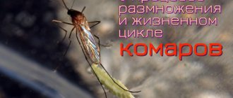
Life cycle of Plasmodium falciparum
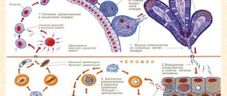
It is a complex process. During evolution, the parasite has adapted to live only in the body of other organisms, choosing two hosts for this: a mosquito and a human. Therefore, malaria infection occurs mainly through an insect bite or directly through the blood. It is believed that the definitive host of Plasmodium falciparum is humans. After all, it is he who displays the symptoms of the disease, and the mosquito does not even notice that it is a carrier. But in fact, it is in the insect’s body that sexual reproduction of the parasite occurs. Therefore, scientists have proven that humans are the intermediate host of malarial plasmodium. What development cycle does the parasite go through?
1. When sucking infected blood, immature germ cells of plasmodium enter the mosquito’s body. It is there that they are fertilized and, having attached themselves to the mosquito’s stomach, begin to divide. The total number of parasite sporozoids can reach hundreds of thousands; a larger number of them are found in the salivary glands of the mosquito. That is why the insect is the main host of the malarial plasmodium.
2. When a mosquito bites, sporozoids enter the human blood. There the parasite goes through two phases of development. It divides in the liver, forming merozoites, and then penetrates red blood cells, where it undergoes asexual reproduction.
The main host of Plasmodium falciparum
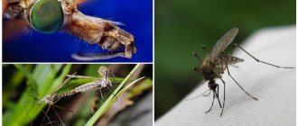
The carrier of the disease is a female mosquito of one species - living in hot climates. This is due to the fact that the development of malarial plasmodium is possible only at temperatures above 16 degrees. And this process is most active at 25-28 degrees. Plasmodium germ cells that enter the mosquito's body can only be fertilized there. Within 10-15 minutes, a zygote is formed, which turns into a sporocyst and attaches to the outer wall of the insect’s stomach. There she begins to divide. Several thousand sporozoids are formed in one sporocyst. And there can be a huge number of such fertilized cells in the mosquito’s body.
Therefore, the number of sporozoids can reach hundreds of thousands. They spread throughout the mosquito's body, accumulating in its salivary glands. This is how the sporozoids of Plasmodium falciparum penetrate into the human blood.
Sources
- Yildiz Y., Yavuz A.Y. Complementary and alternative medicine use in children with asthma. // Complement Ther Clin Pract - 2022 - Vol43 - NNULL - p.101353; PMID:33756219
- Carpagnano G., Sessa F., Scioscia G., Lacedonia D., Foschino MP., Venuti MP., Triggiani AI., Valenzano A., Resta O., Cibelli G., Messina G. Physical Activity as a New Tool to Evaluate the Response to Omalizumab and Mepolizumab in Severe Asthmatic Patients: A Pilot Study. // Front Pharmacol - 2022 - Vol10 - NNULL - p.1630; PMID:32038267
- Papadopoulos NG., Čustović A., Cabana MD., Dell SD., Deschildre A., Hedlin G., Hossny E., Le Souëf P., Matricardi PM., Nieto A., Phipatanakul W., Pitrez PM., Pohunek P., Gavornikova M., Jaumont X., Price D.B. Pediatric asthma: An unmet need for more effective, focused treatments. // Pediatr Allergy Immunol - 2022 - Vol30 - N1 - p.7-16; PMID:30312503
- Yang X., Wang X., Chi M., Zhang M., Shan H., Zhang QH., Zhang J., Shi J., Zhang JZ., Wu RM., Li YL. Osteoprotegerin mediate RANK/RANKL signaling inhibition eases asthma inflammatory reaction by affecting the survival and function of dendritic cells. // Allergol Immunopathol (Madr) - 2022 - Vol47 - N2 - p.179-184; PMID:30292447
- Ortega H., Llanos JP., Lafeuille MH., Duh MS., Germain G., Lejeune D., Sama S., Bell C., Hahn B. Effects of systemic corticosteroids on blood eosinophil counts in asthma: real-world data . // J Asthma - 2022 - Vol56 - N8 - p.808-815; PMID:30130418
- Dilokthornsakul P., Thompson AM, Campbell JD. Treatment patterns and related clinical consequences in adults with asthma. // J Asthma - 2022 - Vol56 - N7 - p.739-747; PMID:29972095
- Müller A., Balatoni I., Csernoch L., Bács Z., Bíró M., Bendíková E., Pesti A., Bácsné Bába É. . // Orv Hetil - 2022 - Vol159 - N27 - p.1103-1112; PMID:29961374
- Zhang J., Fulgar CC., Mar T., Young DE., Zhang Q., Bein KJ., Cui L., Castañeda A., Vogel CFA., Sun X., Li W., Smiley-Jewell S., Zhang Z., Pinkerton KE. TH17-Induced Neutrophils Enhance the Pulmonary Allergic Response Following BALB/c Exposure to House Dust Mite Allergen and Fine Particulate Matter From California and China. // Toxicol Sci - 2022 - Vol164 - N2 - p.627-643; PMID:29846732
- Corren J., Kavati A., Ortiz B., Colby JA., Ruiz K., Maiese BA., Cadarette SM., Panettieri RA. Efficacy and safety of omalizumab in children and adolescents with moderate-to-severe asthma: A systematic literature review. // Allergy Asthma Proc - 2022 - Vol38 - N4 - p.250-263; PMID:28631599
- Krogh N., Rzeppa S., Dyreborg A., Dehnes Y., Hemmersbach P., Backer V., Hostrup M. Terbutaline Accumulates in Blood and Urine after Daily Therapeutic Inhalation. // Med Sci Sports Exerc - 2017 - Vol49 - N6 - p.1236-1243; PMID:28072631
Life of a parasite in the human body
When a mosquito that carries the microorganism bites, malarial plasmodium enters the human blood. The parasite in the body goes through two phases of development: tissue and erythrocyte. It is in the second stage that plasmodium causes the symptoms of malaria.
1. The first phase is called tissue schizogony. Penetrating into the liver cells, the sporozoid begins to divide, forming up to 50 thousand merozoites. They enter the bloodstream, penetrating into red blood cells. This stage does not cause any symptoms and lasts from 5 to 16 days. In some cases, for example, when infected with Plasmodium falciparum, some of the sporozoids remain dormant in the liver, causing a relapse of the disease after six months.
2. When plasmodium leaves the liver into the blood, it penetrates into red blood cells, and the cyclic process of erythrocyte schizogony begins. Feeding on hemoglobin, merozoites develop and divide, forming new cells: asexual schizonts and sexual gametocytes. By destroying the red blood cell, they enter the blood plasma. At this point, the person begins to have an attack of fever. When gametocytes form in a person’s blood, it becomes a source of infection, and when a mosquito bites, plasmodium enters the insect’s body and begins sexual reproduction there.
- 1. Trichomonas (Trichomonas vaginalis) and T. hominis
- 2. Giardia (Lamblia intestinalis)
- 3. Leishmaniae
- 4. Trypanosoma
- 5. General characteristics of the class Sporozoans
- 6. Toxoplasmosis: pathogen, characteristics, development cycle, prevention
- 7. Malarial plasmodium: morphology, development cycle
LECTURE No. 19. Pathogenic flagellates
Of medical importance are those species of flagellates that parasitize the body of humans and animals.
Trypanosoma are the causative agents of African and American sleeping fevers. These flagellates live in the tissues of the human body. Their transmission to the host is carried out transmissibly, i.e. through carriers.
Leishmania (Leishmania) are the causative agents of leishmaniasis, vector-borne diseases with natural focality. Carriers are mosquitoes. Natural reservoirs are rodents, wild and domestic predators.
There are three main forms of diseases caused by Leishmania - cutaneous, visceral and mucocutaneous leishmaniasis.
Lamblia intestinalis is the only species of protozoan that lives in the small intestine. Causes lambliasis. Giardia can penetrate the bile ducts and liver.
1. Trichomonas (Trichomonas vaginalis) and T. hominis
These are the causative agents of trichomoniasis. They live in the reproductive and urinary tracts.
Morphological characteristics of Trichomonas
Trichomonas (class Flagellates) are the causative agents of diseases called trichomoniasis. The human body is inhabited by intestinal and vaginal (urogenital) trichomonas.
Urogenital Trichomonas (Trichomonas vaginalis) is the causative agent of urogenital trichomoniasis. In women, this form lives in the vagina and cervix, in men - in the urethra, bladder and prostate gland. Found in 30–40% of women and 15% of men. The disease is widespread.
The length of the parasite is 15–30 µm. The body shape is pear-shaped. It has 4 flagella, which are located at the anterior end of the body.
There is an undulating membrane that extends to the middle of the body. In the middle of the body there is an axostyle, protruding from the cell at its posterior end in the form of a spike. The core has a characteristic shape: oval, pointed at both ends, reminiscent of a plum pit. The cell contains digestive vacuoles in which leukocytes, erythrocytes and bacteria of the genitourinary flora can be found, on which the urogenital Trichomonas feeds. Does not form cysts.
Infection most often occurs through sexual contact during unprotected sexual contact, as well as through the use of a shared bed and personal hygiene items: towels, washcloths, etc. Non-sterile gynecological instruments and gloves during a gynecological examination can serve as a transmission factor.
This parasite usually does not cause visible harm to the host, but causes chronic inflammation in the genitourinary tract. This occurs due to close contact of the pathogen with the mucous membranes. In this case, epithelial cells are damaged, it desquamates, microinflammatory foci and erosions appear on the surface of the mucous membranes.
In men, the disease can spontaneously recover 1–2 months after infection. Women get sick longer (up to several years).
Diagnostics. Based on the detection of vegetative forms in a smear of discharge from the genitourinary tract.
Prevention – compliance with the rules of personal hygiene, the use of personal protective equipment during sexual intercourse.
Intestinal Trichomonas (Trichomonas hominis) is a small flagellate (length - 5-15 microns) that lives in the large intestine. It has 3–4 flagella, one nucleus, an undulating membrane and an axostyle. Feeds on bacteria of the intestinal flora. The formation of cysts has not been established.
Infection occurs through food and water contaminated with Trichomonas. Once in the intestines, the parasite multiplies quickly and can cause diarrhea. It is also found in the intestines of healthy people, i.e. carriage is possible.
Diagnostics. Based on the detection of vegetative forms in feces.
Prevention.
1. Personal. Compliance with personal hygiene rules, heat treatment of food and water, thorough washing of vegetables and fruits (especially those contaminated with soil).
2. Public. Sanitary arrangement of public places, monitoring of public water supply sources, sanitary and educational work with the population.
2. Giardia (Lamblia intestinalis)
Giardia belongs to the class Flagellates. This is the only protozoan that lives in the human small intestine. Causes a disease called intestinal giardiasis. Most often it affects young children.
It lives in the small intestine, mainly in the duodenum, and can penetrate the bile ducts (intrahepatic and extrahepatic), and from there into the gallbladder and liver tissue. Giardiasis is widespread.
Morphology
The dimensions of the parasite are 10–18 microns. The body shape resembles a pear cut in half. The body is clearly divided into right and left halves. In this regard, all organelles and nuclei are paired. There are 2 lunar-shaped nuclei (in the middle of the body) and 4 pairs of flagella located symmetrically. In the expanded part there is a suction disk, with the help of which the parasite attaches to the villi of the small intestine. There are 2 thin axo-styles running along the body.
Features of the life of Giardia
Giardia is capable of forming cysts, which are excreted in feces and thus spread in the environment. Cysts form in the lower parts of the small intestine.
Mature cysts are oval in shape, contain 4 nuclei and several supporting axostyles. In the external environment, they are quite resistant to adverse conditions and remain viable for several weeks.
Humans become infected by ingesting cysts that get into food or drinking water.
Excision occurs in the small intestine and vegetative forms (trophozoites) are formed. With the help of suction cups they attach to the villi of the small intestine.
Giardia uses nutrients that they capture from the surface of intestinal epithelial cells using pinocytosis. If there is a large number of Giardia in the intestine, they are able to cover fairly large surfaces of the intestinal epithelium.
In this regard, the processes of parietal digestion and absorption of food are significantly disrupted. In addition, the presence of Giardia in the intestines causes inflammation. Penetrating into the bile ducts, they cause inflammation of the gallbladder and disrupt the outflow of bile.
Giardia can occur in apparently healthy people. Then asymptomatic carriage is observed. However, these people are dangerous because they can infect others.
Diagnostics. Based on the detection of cysts in feces. Trophozoites can be detected in the contents of the duodenum obtained by fractional duodenal intubation.
Prevention.
1. Personal. Compliance with personal hygiene rules (such as washing hands before eating and after visiting the toilet, thoroughly washing fruits and vegetables, heat treatment of food and drinking water, etc.).
2. Public. Sanitary improvement of public toilets, catering establishments, sanitary education work with the population.
3. Leishmaniae
Leishmania is a protozoan of the flagellate class. They are causative agents of leishmaniasis - vector-borne diseases with natural focality.
Diseases in humans are caused by several species of this parasite: L. tropica - the causative agent of cutaneous leishmaniasis, L. donovani - the causative agent of visceral leishmaniasis, L. brasiliensis - the causative agent of Brazilian leishmaniasis, L. mexicana - the causative agent of the Central American form of the disease. They all have morphological similarities and the same development cycles.
They exist in two forms: flagellated (leptomonadous, otherwise promastigote) and flagellate (leishmanial, otherwise amastigote).
The leishmanial form is very small (3–5 µm), round. It does not have a flagellum. Lives in the cells of the reticuloendothelial system of humans and some animals (rodents, dogs). The flagellar form is elongated (up to 25 µm), and has a flagellum at the anterior end. Found in the digestive tract of carriers (small mosquitoes of the genus Phlebotomus). These forms can also form in artificial cultures. The natural reservoir is rodents, wild and domestic predators.
Leishmania is widespread in countries with tropical and subtropical climates, on all continents where mosquitoes exist.
In cutaneous leishmaniasis, the lesions are located in the skin. This is the most common form. The disease progresses relatively favorably. Caused by L. tropica, L. mexicana and some biovars of L. brasiliensis. After a mosquito bite, round, non-healing ulcers form on exposed parts of the body. After healing, scars remain. Lifelong immunity. Some forms of L. brasiliensis can migrate through the lymphatic vessels, causing ulcers to form far from the site of the bite.
The mucocutaneous form is caused by the subspecies L. brasiliensis brasiliensis. Leishmania penetrates from the skin through the blood vessels into the mucous membrane of the nasopharynx, larynx, soft palate, and genitals, causing destructive changes in the mucous membranes.
Diagnostics
They take discharge from a skin or mucous ulcer and prepare smears for subsequent microscopy.
The visceral form of the disease is caused by L. donovani. The incubation period is long, and the disease begins several months or years after infection. Children under 12 years of age are more likely to get sick. The disease occurs as a systemic infection. Parasites multiply in tissue macrophages and blood monocytes. Intoxication is very high. Liver function and hematopoiesis are impaired. If left untreated, the disease is fatal.
Diagnostics
A punctate of red bone marrow (during puncture of the sternum) or lymph nodes is obtained, followed by the preparation of a smear or print for microscopy. In stained preparations, the leishmanial form of the parasite is found, both extra- and intracellularly localized. In doubtful cases, the material is inoculated on nutrient media, where the leishmanial form turns into a flagellated one, actively moves and is detected by ordinary microscopy. Biological samples are used (for example, infection of laboratory animals).
Prevention
Control of vectors (mosquitoes), destruction of natural reservoirs, preventive vaccinations.
4. Trypanosoma
The causative agents of trypanosomiasis are trypanosomes (class Flagellates). African trypanosomiasis (sleeping fever) is caused by Trypanosoma brucei gambiensi and T. b. rhodesiense. American trypanosomiasis (Chagas disease) is caused by Trypanosoma cruzi.
The parasite has a curved body, flattened in one plane, pointed on both sides. Dimensions – 15–40 microns. The stages that live in the human body have 1 flagellum, an undulating membrane and a kinetoplast located at the base of the flagellum.
In the human body and other vertebrates, the parasite lives in blood plasma, lymph, lymph nodes, cerebrospinal fluid, the substance of the brain and spinal cord, and serous fluids.
The disease is widespread throughout Africa.
Trypanosomiasis, caused by these parasites, is a typical vector-borne disease with natural focality. The causative agent of trypanosomiasis develops with a change of hosts. The first part of the life cycle takes place in the body of the carrier. Tripanosoma brucei gambiensi is carried by tsetse flies Glossi-na palpalis (lives near human habitation), T. b. rho-desiense, Glossina morsitans (in open savannas). The second part of the life cycle takes place in the body of the final host, which can be large and small cattle, humans, pigs, dogs, rhinoceroses, and antelopes.
When a tsetse fly bites a sick person, the trypanosomes enter the fly's stomach. Here they reproduce and go through several stages. The full development cycle takes 20 days. Flies whose saliva contains trypanosomes in an invasive (meta-cyclic) form can infect humans when they bite.
Without treatment, sleeping sickness can last a long time (up to several years). Patients experience progressive muscle weakness, exhaustion, drowsiness, depression, and mental retardation. Self-healing is possible, but most often without treatment the disease ends in death. Trypanosomiasis caused by T. b. Rhodesiense, is more malignant and ends in death 6–7 months after infection.
Diagnostics
Blood smears and cerebrospinal fluid are examined, and a biopsy of lymph nodes in which pathogens are visible is performed.
Prevention
Vector control, preventive treatment of healthy people in areas of trypanosomiasis, making the body immune to the pathogen.
Trypanosoma cruzi is the causative agent of American trypanosomiasis (Chagas disease). The pathogen is characterized by the ability to live intracellularly. They multiply only in the cells of the myocardium, neuroglia and muscles (in the form of non-flagellar forms), but not in the blood.
Vectors are triatomine bugs. Trypanosomes multiply in their body. After a bite, bedbugs defecate; the pathogen, in its infective stage, enters the wound with feces. The pathogen lives in the tissues of the heart, brain, and muscles. This disease is characterized by myocarditis, hemorrhages in the meninges, and their inflammation.
Diagnostics
Detection of the pathogen in the blood (in the acute period). In a chronic course, infection of laboratory animals occurs.
Prevention
The same as for African trypanosomiasis.
5. General characteristics of the class Sporozoans
About 1,400 sporozoan species are known. All representatives of the class are parasites (or commensals) of humans and animals. Many sporozoans are intracellular parasites. It is these species that have undergone the most profound degeneration in terms of structure: their organization has been simplified to a minimum. They do not have any excretory or digestive organs. Nutrition occurs due to the absorption of food by the entire surface of the body. Waste products are also released through the entire surface of the membrane. There are no respiratory organelles. The common features of all representatives of the class are the absence of any organelles of movement in mature forms, as well as a complex life cycle. Sporozoans are characterized by two variants of the life cycle - with and without the presence of the sexual process. The first version of the cycle includes the stages of asexual reproduction and the sexual process (in the form of copulation and sporogony).
Asexual reproduction is carried out by simple division through mitosis or multiple division (schizogony). In schizogony, multiple divisions of the nucleus occur without cytokinesis. Then the entire cytoplasm is divided into parts, which are separated around the new nuclei. From one cell many daughter cells are formed. Before the sexual process, the formation of male and female reproductive cells - gametes - occurs. They are called gamonts. The different-sex gametes then fuse to form a zygote. It is covered with a dense shell and turns into a cyst, in which sporogony occurs - multiple divisions with the formation of cells (sporozoites). It is at the sporozoite stage that the parasite penetrates the host body. Sporozoans, which are characterized by just such a development cycle, live in the tissues of the internal environment of the human body (for example, malarial plasmodia).
The second version of the life cycle is much simpler and consists of a cyst stage and a trophozoite (an actively feeding and reproducing form of the parasite). This development cycle is found in sporozoans, which live in cavity organs communicating with the external environment.
Basically, sporozoans, parasitic in the body of humans and other vertebrates, live in body tissues. They can affect both humans and many animals (including wild ones). Thus, these are zoo- and anthropozoonotic diseases, the prevention of which is a difficult task. These diseases can be transmitted non-transmissibly (like toxoplasma), i.e., without a specific carrier, or transmissible (like malarial plasmodia), i.e., through carriers.
Diagnosis of diseases caused by protozoa of the class Sporozoans is quite difficult, since parasites can live in various organs and tissues (including deep ones), which reduces the likelihood of their detection. In addition, the severity of the symptoms of the disease is low, since they are not strictly specific.
Toxoplasma gondii is the causative agent of toxoplasmosis. Humans are the intermediate host for this parasite, and the main hosts are cats and other representatives of the feline family.
Malarial plasmodia (Plasmodium) are the causative agents of malaria. Humans are the intermediate host, the final host being mosquitoes of the genus Anopheles.
6. Toxoplasmosis: pathogen, characteristics, development cycle, prevention
The causative agent of toxoplasmosis is a representative of the class Toxoplasma gondii. It affects a huge number of animal species, as well as humans.
The parasite, localized in cells, has the shape of a crescent, one end of which is pointed and the other is rounded. At the center of the cell is the nucleus. At the pointed end there is a structure similar to a suction cup - a conoid. It serves for fixation and penetration into host cells.
The life cycle is typical for sporozoans. There is an alternation of asexual and sexual reproduction - schizogony, gametogenesis and sporogony. The definitive hosts of the parasite are cats and other representatives of the feline family. They receive the pathogen by eating the meat of sick animals (rodents, birds) or contaminated meat of large herbivores. In cat intestinal cells, parasites first reproduce by schizogony, producing many daughter cells. Next, gametogenesis occurs and gametes are formed. After their copulation, oocysts are formed, which are released into the external environment. Sporogony occurs under the cyst shell and many sporozoites are formed.
Sporocysts with sporozoites enter the body of an intermediate host - humans, birds, many mammals and even some reptiles.
Once in the cells of most organs, Toxoplasma begins to actively multiply (multiple fission). As a result, a huge number of pathogens appear under the membrane of one cell (a pseudocyst is formed). When one cell is destroyed, many pathogens come out of it and penetrate other cells. Other groups of Toxoplasma in the host cells become covered with a thick membrane, forming a cyst. Toxoplasma can persist in this state for a long time. They are not released into the environment. The development cycle is completed when cats eat contaminated meat from intermediate hosts.
In the body of a sick person, toxoplasma is found in the cells of the brain, liver, spleen, lymph nodes and muscles. A person, as an intermediate host, can acquire toxoplasma by eating the meat of infected animals, through damaged skin and mucous membranes when caring for sick animals, when processing infected meat or skins, transplacentally (toxoplasma can pass through a healthy placenta), during medical procedures - donor transfusion blood and its preparations, transplantation of donor organs while taking immunosuppressants (suppressing the body’s natural defenses).
In most cases, asymptomatic parasite carriage or a chronic course without characteristic symptoms are observed (if the parasites have low pathogenicity). In rare cases, the disease is acute: with a rise in temperature, enlargement of peripheral lymph nodes, the appearance of a rash and manifestations of general intoxication. This is determined by the individual sensitivity of the body and the routes of penetration of the parasite.
Prevention
Heat treatment of food products of animal origin, sanitary control in slaughterhouses and meat processing plants, exclusion of contacts of pregnant women and children with pets.
7. Malarial plasmodium: morphology, development cycle
Malarial plasmodia belong to the class Plasmodium and are the causative agents of malaria. The following types of plasmodia parasitize the human body: P. vivax - the causative agent of tertian malaria, P. malariae - the causative agent of tetanus malaria, P. falciparum - the causative agent of tropical malaria, P. ovale - the causative agent of ovalemalaria, close to tertian malaria (found only in Central Africa). The first three species are common in tropical and subtropical countries. All types of plasmodia have similar structural features and life cycle; the only difference is in certain details of morphology and some features of the cycle.
The life cycle is typical for sporozoans and consists of asexual reproduction (schizogony), sexual process and sporogony.
Malaria is a typical anthroponotic vector-borne disease. The carriers are mosquitoes of the genus Anopheles (they are also the definitive hosts). The intermediate host is only human.
Human infection occurs through the bite of a mosquito whose saliva contains plasmodia at the sporozoite stage. They penetrate the blood, with a current that ends up in the liver tissue. Tissue (pre-erythrocytic) schizogony occurs here. It corresponds to the incubation period of the disease. In liver cells, tissue schizonts develop from sporozoites, which increase in size and begin to divide schizogony into thousands of daughter individuals. In this case, the liver cells are destroyed, and parasites at the merozoite stage enter the blood. They invade erythrocytes, in which erythrocyte schizogony occurs. The parasite absorbs hemoglobin from blood cells, grows and multiplies by schizogony. Moreover, each plasmodium produces from 8 to 24 merozoites. Hemoglobin consists of an inorganic iron-containing part (heme) and protein (globin). The parasite feeds on globin. When the affected red blood cell bursts, the parasite enters the bloodstream and heme enters the blood plasma. Free heme is a powerful poison. It is its entry into the blood that causes terrible attacks of malarial fever. The patient's body temperature rises so high that in the old days, malaria infection was used as a treatment for syphilis (Spanish scabies): treponema cannot withstand such temperatures. The development of plasmodia in erythrocytes goes through four stages: ring (trophozoite), amoeboid schizont, fragmentation (morula formation) and (for some parasites) gametocyte formation. When an erythrocyte is destroyed, merozoites enter the blood plasma, and from there into new erythrocytes. The cycle of erythrocyte schizogony is repeated many times. The growth of the trophozoite in the erythrocyte takes time, constant for each type of plasmodium. An attack of fever is timed to coincide with the release of parasites into the blood plasma and is repeated every 3 or 4 days, although with a long-term disease the alternation of periods may be unclear.
From some of the merozoites in erythrocytes, immature ha-monts are formed, which are an invasive stage for the mosquito. When a mosquito bites a sick person, the gamonts enter the mosquito's stomach, where they form mature gametes. After fertilization, a motile zygote (ookinete) is formed, which penetrates under the epithelium of the mosquito’s stomach. Here it increases in size, becomes covered with a dense shell, and an oocyst is formed. Multiple divisions occur inside it, during which a huge number of sporozoites are formed. Then the oocyst shell bursts, and plasmodia penetrate through the bloodstream into all tissues of the mosquito. Most of them accumulate in his salivary glands. Therefore, when a mosquito bites, sporozoites can enter the human body.
Thus, in humans, plasmodium reproduces only asexually - by schizogony. Humans are an intermediate host for the parasite. The sexual process takes place in the mosquito's body - the formation of a zygote, many sporozoites are formed (sporogony occurs). The mosquito is the final host, and it is also the carrier.
Malaria: pathogenic significance, diagnosis, prevention.
Malaria is a serious disease characterized by periodic, debilitating attacks of fever with chills and heavy sweating. When a large number of merozoites leave erythrocytes, many toxic waste products of the parasite itself and breakdown products of hemoglobin, which the plasmodium feeds on, are released into the blood plasma. When exposed to them on the body, severe intoxication occurs, which manifests itself in a sharp paroxysmal increase in body temperature, the appearance of chills, headaches and muscle pain, and severe weakness. Temperatures can reach significant levels (40–41 °C). These attacks occur acutely and last on average 1.5–2 hours. This is followed by thirst, dry mouth, and a feeling of heat. After a few hours, the temperature drops to normal levels, all symptoms stop, and patients fall asleep. In general, the entire attack lasts from 6 to 12 hours. There are differences in the intervals between attacks in different types of malaria. With three-day and oval malaria, attacks are repeated every 48 hours. Their number can reach 10–15, after which they stop, as the body begins to produce antibodies against the pathogen. Parasites can still be detected in the blood, so a person becomes a carrier of the parasite and poses a danger to others.
In malaria caused by P. malariae, the intervals between attacks are 72 hours. Asymptomatic carriage is common.
With tropical malaria, at the onset of the disease, the intervals between attacks can be different, but then they are repeated every 24 hours. With this type of malaria, there is a high risk of death due to complications from the central nervous system or kidneys. Tropical malaria is especially dangerous for representatives of the Caucasian race.
A person can become infected with malaria not only through the bite of an infected mosquito. Infection is also possible through blood transfusion (transfusion) of infected donor blood. Most often, this method of infection occurs with four-day malaria, since there are few schizonts in erythrocytes, they may not be detected when testing the blood of donors.
Diagnostics
It is possible only during the period of erythrocyte schizogony, when the pathogen can be detected in the blood. Plasmodium, which has recently penetrated into an erythrocyte, has the appearance of a ring. The cytoplasm in it, in the form of a rim, surrounds a large vacuole. The core is shifted to the edge.
Gradually the parasite grows, and pseudopods appear (in the amoeboid schizont).
It occupies almost the entire red blood cell. Next, fragmentation of the schizont occurs: in the deformed erythrocyte, many merozoites are found, each of which contains a nucleus. In addition to asexual forms, gametocytes can also be found in erythrocytes. They are larger and do not have pseudopods or vacuoles.
Prevention
Identification and treatment of all patients with malaria (elimination of the source of mosquito infestation) and destruction of mosquitoes (elimination of vectors) using special insecticides and reclamation work (draining swamps).
When traveling to areas unfavorable for malaria, you should take prophylactic antimalarial drugs and protect yourself from mosquito bites (use mosquito nets, apply repellents to the skin).
Table of contents
buy female pathogen
Development of the parasite in human blood
Why is malarial plasmodium so dangerous for humans? Its reproduction in human blood leads to the destruction of red blood cells. In these blood cells, the parasite undergoes a number of changes: merozoites pass into the trophozoid stage, which feeds on hemoglobin and grows rapidly, then schizonts are formed. They reproduce asexually and destroy red blood cells. At the same time, foreign protein, potassium salts, remnants of destroyed blood cells and metabolic products of the parasite enter the blood.
In addition to attacks of fever, this process causes disruption of the liver and spleen, leading to the growth of connective tissue in them. The breakdown of red blood cells also causes anemia. The brain is often affected, which occurs due to increased capillary permeability.
Diagnostics
If a person develops the first symptoms and is at risk.
The first thing he should do is take a blood test and conduct a biochemical analysis, which will reveal anemia (low hemoglobin and red blood cells), low platelet levels and an excessive white blood cell count.
The second test is to determine malarial plasmodia in the blood (detailed examination of a drop of blood). If detected, specific therapy is prescribed.
Features of malaria
This disease, called “swamp fever,” has been known since ancient times. It was widespread in all tropical countries. Only in the 17th century did they begin to successfully treat the disease using cinchona bark. And at the end of the 19th century, the causative agent of malaria was discovered. It was only in the mid-20th century that the life cycle of malarial plasmodium was studied in detail and effective drugs were created to treat the disease. Despite this, more than 300 million people still fall ill with malaria every year in hot countries. About two million cases are fatal. This disease is characterized by four stages:
- Incubation period during which weakness and headache are observed.
- Second stage, febrile. It begins with severe chills. At this time, the pulse quickens, the pressure rises, and the patient cannot warm up. After 1-3 hours, the temperature rises significantly - up to 41 degrees, delirium, convulsions and headache are observed. After this, the temperature drops, which is accompanied by heavy sweating.
— After 10-12 attacks, the infection subsides and a secondary latent period is observed.
- If the treatment was incorrect, then after a few months the disease relapses. It can also occur in case of infection with one of the types of plasmodium that causes tropical malaria. Some of the sporozoids of this parasite remain in the human liver in a dormant state.
What is the danger of the disease
In addition to recurring periods of fever, malaria causes disruption of all human systems and organs. Myocardial dystrophy, neuritis, migraines, nephritis, thrombocytopenia and anemia may develop. The liver and kidneys are severely affected. In some cases, the disease is chronic and difficult to treat. Children are especially susceptible to infection; the mortality rate among them is quite high. Until now, the disease is very common in Africa, Australia, South America, on the coast of the Red and Mediterranean Seas, in Southeast Asia and India.
Treatment and prevention of malaria
Even in modern society it is not possible to cope with this disease. After all, the main host of the malarial plasmodium is the mosquito. And in tropical countries, especially with a humid climate, there are a lot of these insects. Therefore, the most important thing in preventing the disease is to protect yourself from insect bites. To do this, you can use mosquito nets or repellents. To treat the disease in case of infection, many drugs are now used: Chlorozin, Mefloquine, Primaquine, Akrikhin and Quinine, known since the 19th century. They not only quickly relieve the symptoms of an incipient attack, but also completely destroy the parasite. People living in tropical areas and having had the disease acquire immunity to this type of pathogen, but can be carriers of the infection. And those who are planning to travel to such countries are advised to constantly take anti-malarial drugs.
Comments 1
| -1 quantum 01/19/2021 11:04 [Material] Maybe it will be useful to someone. Mom tried for a long time to explain to me the seeds of which plant the doctor brewed with them during the war to cure malaria. I finally found it and she confirmed that it was it. True, she was little and only remembers that they brewed it together with the transparent shell in a teapot and drank it like tea. He doesn’t remember how much or how much water. But he says it saved them. The plant is called Lunnik, we used to have a lot of it as an ornamental plant, but also medicinal. But I couldn’t find anywhere that it treats malaria. But my mother claims that everyone who knew was saved by him. Something radical.ru doesn’t work.... here’s the link then, there’s a picture https://clck.ru/SrHaZ |
Add comments can only registered users. [Registration | Entrance ]