General characteristics of arachnids
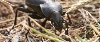
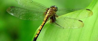
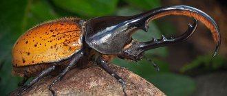
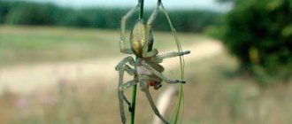
Arachnids are a large group of exclusively terrestrial chelicerates, numbering over 36,000 species. Coming onto land and settling along it, arachnids acquired various adaptations to very diverse living conditions. We will find arachnids in the field, forest, garden and vegetable garden, in courtyards, in human dwellings, etc. There are many parasitic forms among them. Representatives of various orders of arachnids - scorpion, telephon, false scorpion, harvestman, salpuga, spider and tick.
External structure
Like typical chelicerates, the body of the vast majority of arachnids consists of a fused cephalothorax, bearing six pairs of limbs, and an abdomen. The abdomen, unlike horseshoe crabs, does not bear real limbs. Only their rudiments or limbs, transformed into special organs, are found.
Antennae or antennules are absent. The eyes are simple. The first pair of limbs of the cephalothorax is located in front of the mouth. These are short chelicerae, consisting of 2-3 segments, ending in a claw, hook or stylet. Chelicerae are homologous to the second antennae of crustaceans. Behind the mouth there is a second pair of limbs - the pedipalps. Their bases have chewing processes, and the remaining segments can serve as tentacles. Pedipalps can turn into walking legs or food-grasping organs - powerful claws (scorpions, pseudoscorpions). All arachnids typically feed on liquid food, so the anterior section of the digestive system is a sucking apparatus.
In connection with the arrival on land, arachnids transformed some organ systems of the primary aquatic chelicerates and new ones emerged. In some groups, both old and newly acquired organs exist simultaneously. Thus, the respiratory organs of arachnids are the lungs, located in pairs on the abdominal segments. Their origin and development prove that they are modified gill legs of aquatic chelicerates. The new respiratory organs of arachnids are tracheas - blind invaginations of the outer integument.
The excretory organs are also dual in nature. They are represented by coxal glands (coelomoducts) that are more ancient in origin and newly emerged Malpighian vessels.
The differences between representatives of arachnid orders lie in the degree of segmentation of the body, primarily the abdomen, and in the specialization of the cephalothorax limbs, adapted to perform various functions. The body of scorpions is most strongly segmented. It consists of a small fused cephalothorax and abdomen, represented by 12 segments, of which 6 wider segments constitute the anterior belly, or mesosoma, and the remaining 6 narrower ones constitute the posterior abdominal, or metasoma. Attention should be paid to the similarity in the body dismemberment of scorpions and the extinct gigantic crustacean scorpions. In both cases, the metasoma is represented by six segments. In other groups of arachnids, the posterior part of the abdomen, the metasoma, contracts, and the abdomen shortens. In terms of the degree of dissection of the abdomen, flagipods and pseudoscorpions are close to scorpions, in which, however, the abdomen is not externally divided into anterior and posterior abdominals. In some respects, salpugs are even more dissected animals than scorpions. In addition to the articulated abdomen, which has 10 segments, salpugs have two free thoracic segments that are not part of the fused head. The articulated abdomen of harvestmen also consists of 10 segments, which are not separated by a deep constriction from the cephalothorax, as in real spiders. In arthroplasty spiders (four-lunged) the abdomen consists of 11 segments, and in higher spiders it consists of 6, while the abdominal segments are completely fused. In ticks, the number of abdominal segments is reduced to 7, and in some - to 4-2. Moreover, in most ticks not only are all segments of the abdomen fused, but it is also impossible to distinguish between the main sections - the cephalothorax and abdomen, which form one whole. Thus, it is obvious that the evolution of various orders of arachnids went in the direction of reducing the number of abdominal segments and their fusion, reducing the degree of overall body dismemberment.
In representatives of various orders, the chelicerae and pedipalps have undergone the greatest changes, and the least changed are the four pairs of walking legs, which have turned into a jointed leg ending in a tarsus with claws.
In scorpions, pseudoscorpions and harvestmen, the chelicerae end in small claws. They play the role of the upper jaws, and, in addition, animals hold prey with them. In salpugs, the chelicerae have turned into powerful claws, adapted for grasping and killing prey. In real spiders, the chelicerae are claw-shaped and consist of two segments. The main segment is very swollen, and the second has a claw-like shape. Near its pointed end, the duct of the poisonous gland opens, located at the base of the chelicera. In a calm state, this segment is applied to the main segment and partially fits into a special groove. With two chelicerae, spiders grab and kill prey, releasing the secretion of the poisonous gland into the wound. Finally, in mites, chelicerae and pedipalps form a piercing-sucking (dog tick, etc.) or gnawing-sucking (scabies mite, barn mite, etc.) oral apparatus.
The second pair of limbs - pedipalps - in salpugs differ little from walking legs, and in scorpions and pseudoscorpions they have turned into grasping organs - claws. In female spiders, the pedipalps play the role of jaws, since they have a chewing plate at the base, and at the same time they are oral tentacles. Male spiders have a swelling on the last segment of the pedipalp, which is a device for fertilizing females. During the breeding season, a special pear-shaped appendage develops on this segment with an elongated end, on which there is an opening leading into a narrow canal, ending inside this organ with an expanded ampulla. Using this device, male spiders collect sperm inside the ampoule and, during mating, inject it into the genital opening of the female.
Abdominal limbs, as such, are absent in all arachnids. However, some of them have survived in a greatly modified form. The rudiments of the abdominal limbs are located only on the mesosoma (anterior six segments). The most complete set of them is preserved in Scorpios. On their first abdominal segment, on which the genital opening is located in all arachnids, there are small genital operculums, and on the second segment there are special comb-like appendages of unknown purpose. The next four segments contain a pair of pulmonary sacs. Four-lunged spiders and flagellated spiders each have two pairs of lungs on the first two abdominal segments; in two-lunged spiders there is one pair of lungs (on the first segment), and on the second, tracheas develop instead of lungs (not connected to the limbs). All spiders develop arachnoid warts on the third and fourth segments - transformed abdominal limbs of these segments. Some groups of small arachnids (part of mites) retain rudiments of abdominal limbs on the first three segments, the so-called coxal organs.
Integuments and skin glands
The body of arachnids is covered with a chitinous cuticle, which is secreted by a layer of flat cells of the hypodermis. In most forms, chitin is poorly developed and the integument is so thin that it wrinkles when dry. Only in some arachnids (scorpions) the chitinous cover is more dense, as it contains calcium carbonate.
Skin (hypodermal) formations include various glands: poisonous, arachnoid, odorous glands of harvestmen, frontal and anal glands of flagellates, etc. Not all arachnids are poisonous. Venom glands are found only in scorpions, spiders, some pseudoscorpions and some ticks. In scorpions, the posterior abdomen ends in a curved caudal spine. At the base of this needle there is a pair of sac-like glands that secrete a poisonous secretion. At the very end of the needle, the openings of the ducts of these glands are placed. Scorpios use this device in a unique way. Having grabbed the prey with the claws of the pedipalps, the scorpion bends its posterior abdomen onto its back and strikes the victim with a needle, from which it releases poison into the wound. In spiders, venom glands are located at the base of the cholicerae, and their ducts open on the claw of the chelicerae.
Arachnoid glands are found mainly in representatives of the order spiders. Thus, the female cross spider (Araneus diadematus) has up to 1000 arachnoid glands of various structures in its abdomen. Their ducts open with tiny holes at the ends of special chitinous cones, which are located on the arachnoid warts and partly on the abdomen near them. Most spiders have 3 pairs of arachnoid warts, but only two of them are formed from the abdominal legs. In some tropical spiders they are multi-segmented.
Pseudoscorpions and spider mites also have arachnoid glands, but they are located in the chelicerae in the former and in the pedipalps in the latter.
Digestive system
Most arachnids are predators, some (ticks) are parasites of animals and plants. Arachnids feed only on semi-liquid food. A number of features in the structure of the digestive apparatus are associated with this method of nutrition. Either completely liquefied food or food containing only very small solid particles is sucked into the intestines of arachnids.
The digestive system consists of three main sections - the foregut, midgut and hindgut.
The foregut with its glands is an organ adapted for liquefying and absorbing food. In spiders, the mouth leads into the pharynx, which is followed by a thin esophagus, which flows into a sucking stomach, driven by muscles running from it to the dorsal integument of the cephalothorax. These three sections (pharynx, esophagus, sucking stomach) are parts of the anterior ectodermic gut and are lined from the inside with chitin. The ducts of the salivary glands open into the pharynx, secreting a secretion that dissolves proteins. Having pierced the integument of the prey, the spider lets saliva into the wound, which dissolves the tissues of the victim, and then sucks out the semi-liquid food. From the sucking stomach begins the endodermic midgut, in which food is digested and absorbed.
The midgut, located in the cephalothorax, forms five pairs of blind glandular projections extending forward to the head end and the bases of the walking legs. Blind outgrowths of the midgut are very characteristic of many arachnids: ticks, harvestmen, etc. They increase the capacity of the intestine and its absorption capacity. In the abdomen, the ducts of the highly developed paired liver flow into the midgut. The liver is a derivative of the midgut. It consists of many thin tubes that not only secrete digestive enzymes, but are also capable of digesting and absorbing nutrients. Intracellular digestion can occur in liver cells. Next, the midgut forms an expanded section, the so-called rectal sac or cloaca, into which the excretory organs, the Malpighian vessels, open. From the rectal sac comes the ectodermic hindgut (rectum), ending in the anus.
The digestive system of other arachnids varies in detail, but is generally structured similarly.
Respiratory system
Due to their land-based lifestyle, arachnids breathe atmospheric air. The respiratory organs of arachnids can be the lungs and trachea. At the same time, it is curious that some arachnids (scorpions, flagellated and four-legged spiders) have only lungs, others (false scorpions, salpugs, harvestmen, and partly ticks) have only tracheas, and finally, still others (most spiders) have both lungs and tracheas.
Four pairs of lungs in scorpions are located on the 3rd-6th segments of the anterior abdomen. On the ventral side, 4 pairs of slit-like openings, stigmata, leading to the lungs are clearly visible. The arachnid lung is a sac-like organ lying on the underside of the abdominal segments. The stigma leads into the lung cavity, which in the anterior part of the pulmonary sac is blocked by plates lying one above the other, which are outgrowths of the lung wall. Between them there are narrow cavities into which air enters. Blood circulates inside the pulmonary plates, and thus an exchange of gases occurs between the blood and the air filling the lungs. Most spiders have one pair of lungs (two-lunged spiders), some have two pairs (four-lunged spiders).
Comparison of the structure of the lung with the structure of the abdominal limbs and gills of horseshoe crabs indicates their great similarity. The position of the lungs on the underside of the abdomen, in the places where the abdominal limbs would be, increases this resemblance. Data from comparative anatomy and embryology fully support the assumption that the lungs of arachnids were formed from the gill legs of fossil merostomes. The transformation of an abdominal limb with gills into a lung can be imagined as follows. A depression formed in the abdominal wall of the body, to which the gills were adjacent, and the lamellar limb grew attached to the integument on the sides. The cavity thus formed communicated with the external environment in the rear part through a narrow, slit-like opening. From the gill filaments, attached only at the wide base to the limb, pulmonary plates with their rather complex structure were formed.
In most arachnids, the trachea serves as the respiratory organ (salpugs, harvestmen, etc.), and in bipulmonary spiders, the trachea exists along with the lungs. The tracheae begin with spiracles (stigmas), usually on the underside of the abdomen. The spiracles can be from one unpaired (in some spiders) to three pairs (in salpugs). The spider's spiracle is located on the abdomen just in front of the arachnoid warts. It leads into two pairs of tracheal tubes, lined from the inside with a thin layer of chitin, which in some arachnids (salpugs, harvestmen and some spiders) forms spiral thread-like thickenings that do not allow the tubes to collapse.
In salpugs, harvestmen and other arachnids, in which the trachea is the only respiratory organ, they form a very complex system of branching tubes that penetrate into all parts of the body and limbs. Some small arachnids lack special respiratory organs; they breathe over the entire surface of the body (a number of species of mites, etc.).
Circulatory system
The circulatory system of arachnids exhibits a metameric structure. Scorpions and most flagipes have a long, tubular heart bearing seven pairs of ostia. In spiders, the number of pairs of ostia is reduced to five or even two. In other arachnids the heart is shorter, and in ticks it is a small vesicle.
Arterial vessels extend forward, backward and to the sides from the heart, and the degree of development and branching of arterial vessels is very different and is directly dependent on the structure of the respiratory organs. Scorpions, which have lungs localized in a specific location, and spiders, whose tracheas are slightly branched, have the most highly developed system of arterial vessels. In salpugs, harvestmen and other forms that breathe by trachea, the system of blood vessels is poorly developed and sometimes absent. This is explained by the fact that with a sufficiently strong branching of the trachea, the exchange of gases occurs directly between the trachea and the tissues of the animal and the blood takes almost no part in the transportation of gases. This is a very interesting example of correlation in the development of various organ systems, even more pronounced in insects.
The degree of development of the circulatory system also depends on the size of the animal. In ticks it is the least developed: some ticks have only a bladder-shaped heart, while others do not have it.
Excretory system
The main excretory organs in arachnids are completely new organs associated with the intestines - the Malpighian vessels. They are one or two pairs of thin tubes, more or less branched and located on the abdomen. These tubes are protrusions of the midgut, i.e. they are of endoderm origin. The Malpighian vessels, blindly closed at the free end, open into the rectal bladder, or cloaca, the last section of the midgut. Guanine, the main excretion product of arachnids, accumulates in their lumens.
Along with the Malpighian vessels, arachnids also have other excretory organs - coxal glands. There may be one or two pairs. They open outward most often at the base of the first and third pairs of walking legs. Typically, the coxal glands consist of a coelomic sac, a nephridial canal, sometimes expanding to form the bladder, and an excretory opening. These organs are apparently homologous to the coelomoducts of annelids and correspond to the coxal glands of horseshoe crabs. In adult arachnids, the coxal glands are usually reduced and do not function; they are replaced by Malpighian vessels.
Nervous system and sensory organs
The nervous system of arachnids is represented by the ventral nerve cord typical of all arthropods. Arachnids are characterized by a significant concentration and fusion of groups of nerve ganglia. The lowest degree of convergence and fusion of ganglia is observed in scorpions. They have a paired suprapharyngeal ganglion (brain), connected by connectives to the cephalothoracic ganglion mass innervating the limbs (2-6 pairs). Next come the seven ganglia of the ventral nerve cord. In salpugas, flageopods and pseudoscorpions, only one of the abdominal ganglia remains free, and the rest join the general ganglion mass. In spiders, all ganglia of the ventral nerve chain form a single subpharyngeal ganglion. In ticks, fusion of the subpharyngeal node is also observed with the brain.
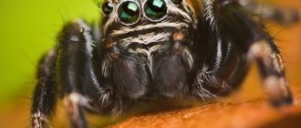
The sense organs include the organs of touch and vision. The organs of touch are the hairs that cover the limbs, especially the pedipalps. The eyes of arachnids are simple (not compounded), usually several pairs. Spiders have 8 eyes located on their heads in two rows.
Sex organs and reproduction
Arachnids are dioecious, and sexual dimorphism can be quite pronounced (in spiders and ticks). In spiders, males are often much smaller than females, and their pedipalps are transformed into a copulatory apparatus.
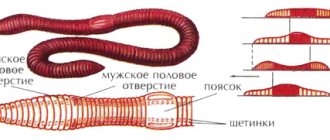
The genital organs of all arachnids consist of paired glands or an unpaired gland that bears traces of the fusion of paired glands. Females have an unpaired gland in the form of a “frame with crossbars” and paired oviducts. Males have paired testes with characteristic crossbars and a copulatory apparatus.
Female spiders have paired spermatic receptacles that open with independent openings in front of the unpaired genital opening on the first abdominal segment. In addition, each of them communicates through a special canal with the uterus, formed by the fusion of the terminal sections of the oviducts.
Using the process of the copulatory apparatus of the pedipalps, spiders inject sperm into the spermatheca of females through their external openings. From there, the sperm enters the uterus, where fertilization occurs.
Partnerogenesis is characterized by ticks. Some species of scorpions are viviparous, and the development of fertilized eggs occurs in the ovaries. Newborn scorpions do not leave their mother, and she carries them on her back for some time.
Development
The development of fertilized eggs in most arachnids is direct. Only in ticks, due to the small size of the eggs, development occurs with metamorphosis. Eggs in most cases are rich in yolk, and crushing is either superficial (spiders, harvestmen, salpugs, mites) or discoidal (oviparous scorpiopes).
In viviparous scorpions, the embryos developing in the mother's ovary consume protein substances secreted by the female's organs. Therefore, despite the small supply of yolk in the eggs of viviparous scorpions, they are characterized by complete crushing.
During embryonic development, arachnids develop a larger number of segments than are present in adult forms. The rudiments of abdominal limbs appear on the abdominal segments, which are subsequently reduced or transformed into other organs.
Limb functions
Arachnids use their limbs for more than just locomotion. In addition, in some species they serve:
- for digging soil;
- in the role of tactile organs;
- to hold food;
- as protection;
- in females to maintain the cocoon;
- for web construction.
Do you know how many pairs of legs are involved in locomotion? It turns out that the first and third pairs of legs on one side of the body move simultaneously with the second and fourth pairs on the other side. The forelimbs do the pulling, and the hind limbs do the pushing.
Phylogeny of arachnids
A number of facts were given above on the basis of which one can imagine the origin of arachnids and the phylogenetic relationships between the orders of this class.
There is no doubt that the terrestrial chelicerates - arachnids - are related to the aquatic chelicerates - crustaceans, and through them with a very ancient and even more primitive group - trilobites. Thus, the evolution of this branch of arthropods proceeded from the most homonomous in segmentation forms, as evidenced by trilobites, to increasingly heteronomous animals.
Of the scientific-like species, the most primitive and ancient group are scorpions, the study of which provides a lot for understanding the evolution of arachnids. Within a class, the evolution of certain groups led to a greater or lesser fusion of abdominal segments, to a greater development of the tracheal system, replacing the more ancient respiratory organs - the lungs, and, finally, to the development of special adaptations characteristic of representatives of individual orders.
Among true spiders, the four-legged spiders are undoubtedly the more primitive group. Two pairs of lungs, the absence of tracheae, the presence of two pairs of coxal glands, and some of them have a segmented abdomen - all these signs indicate their greater primitiveness compared to the group of two-lunged spiders.
Ticks are interesting because their evolution went in the direction of extreme specialization, which is associated with the transition to temporary or permanent parasitism.
Foot sizes
The length, thickness and other dimensions of the legs depend directly on the animal’s lifestyle. Both the first and fourth pair of legs can be the longest. The second pair is always slightly smaller than the first. The third pair is the shortest. In some species, due to the fact that the first two pairs catch and hold prey, the front two pairs are longer than the back ones.
In terms of their structure, the front legs are much thicker than the other limbs. The hips are club-shaped. The paws are attached to the heel, forming an angle with the axis of the leg itself. This structure in some species allows the front pair to be used as tactile organs. They use the other three pairs to move.
Neighborhood with people and interesting facts
Some species of spiders live in close proximity to people. They live where there is sufficient food. Arthropods often eat insects. Typically the following types are found at home:
- Haymaker. Its body size reaches 12 mm, but its paws can be 5 times longer. It mainly weaves webs on windows, which is why it is also called window weaving.
- Tramp. They appear randomly in residential buildings. This species does not spin webs, but hunts on the move.
- Brownie. Body length varies from 6 to 12 mm. Yellow color. They create a funnel-shaped web.
Spiders usually appear when there is a high level of humidity in the house or when there are a large number of different insects. In this case, they multiply quickly and can take over the entire house.
Among the interesting facts about arthropods are the following:
- There is a subspecies in the arachnid family, individuals of which are called phrynes. They are not able to weave webs and have large legs designed for hunting.
- Spiders do not have a skeleton. They only have an outer hard shell, so they can't get too big. Otherwise, the arachnid would have died under its own weight.
- The web is several tens of times stronger than steel.
- All types of spiders make a round web, and only the gladiator makes a square one.
Many people are terrified of spiders. But in fact, there are no serious reasons for this, especially in Russian latitudes. Arachnids are considered the most unusual animals that live on Earth. They received such fame due to the features given by nature.
Features of digestion
The structure of arachnids is adapted to life in a terrestrial environment. The most common spiders are haymakers, cross spiders and jumping spiders. According to their lifestyle, methods of hunting and feeding, arachnids are divided into the following orders:
- spiders;
- scorpions;
- ticks.
The structure of arachnid organs does not allow them to take solid food. Using venom, the spider kills prey and then injects a proteolytic secretion into it. This is the first stage from which the digestion process begins.
The peculiarity of the spider’s digestive system lies in the external or extraintestinal method of processing food.
Internal structure
A distinctive feature of arachnids is that they do not have the usual circulatory system. Instead of blood, they circulate lymph. The heart includes three or four ostia - special openings - lymph moves through them, redirecting to different parts of the body. It fills the space between individual organs, that is, it pours into the cavities. The lymph then enters the pericardial part, and from there it goes back to the heart. Lymph provides all cells of the body with oxygen and removes waste products from the body.
The respiratory system is represented by tracheas and pulmonary sacs, which are shaped like plates. The spiracles are protected by peculiar plugs. They open at the moment of inhalation.
The nervous system is developed at a fairly high level. The nodes are located in the cephalothorax. From them, nerve endings extend to all organs of the arachnid. These nodes are called the animal's brain. Spiders have a large number of nerve cells. This is due to the fact that the brain occupies approximately 30% of the volume of the cephalothorax.
Description of ticks
In accordance with modern taxonomy, the class of arachnids is divided into 9 orders. The most numerous are spiders, ticks and scorpions.
From May to early July, ticks lead the most active lifestyle. During this time, they reproduce and look for food.
The body consists of the abdomen and cephalothorax, and the head includes chelicerae and legs. All departments are fused. What distinguishes spider mites from other representatives is their indirect development cycle. First, a larva hatches from the egg and has 3 pairs of walking legs, but after some time they transform into 4 pairs.
Ticks live in forests, soil, salt and fresh water bodies, on plants, and also parasitize animals and humans. All of these habitats are excellent for nutrition and life.
Many species pose a danger to humans and domestic animals, as they are carriers of serious diseases. They transmit encephalitis, piroplasmosis, and relapsing fever .
The first manifestation of scabies may be the appearance of scabs on the skin and the appearance of severe itching. Representatives of this species gnaw holes under the skin and lay eggs there. After some time, sexually mature individuals hatch. It is very difficult to immediately determine the cause, since the size of the itching varies between 0.2-0.5 mm.
Acne gland parasitizes in hair follicles and sebaceous glands. Because of this parasite, hair falls out and ulcers appear on the skin.
Gall and barn mites do not parasitize humans, but live in pillows, blankets, mattresses, and cracks in the floor. They find dead organic matter for food. But their waste products enter the human body through the respiratory tract, which can lead to severe allergic reactions.