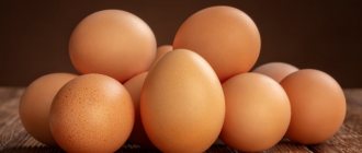
At first glance, an egg, such a common food product for people, has a very complex structure that is difficult to imagine. Even the most insignificant element, it would seem, is called upon to perform important functions in the process of giving birth to a chick. The article examines the detailed structure of an egg laid by a chicken.
Chemical components of a chicken egg
According to its chemical characteristics, a chicken egg is a valuable set of elements. The space enclosed by the shell includes all the required substances necessary for the development of a young organism. The human body absorbs 97% of a bird’s egg, while it receives many amino acids and vitamins A, B, E.
Protein composition
Generally speaking, the composition of the protein in a bird's egg is clearly reflected in its name. In addition to moisture, protein contains many proteins of animal origin:
- Ovoglobulins – about 2%.
- Ovalbumin (reserve for embryo formation) – approximately 54%.
- Highly viscous glycoproteins – up to 3.5%.
- Ovotransferrin (has an antibacterial effect) – up to 13%.
- Lysozyme (an enzyme together with ovotransferrin helps to increase antibacterial properties) - no less than 3.4%.
Egg whites also contain ovomucoid, a compound that provokes allergic reactions in the human body. Therefore, one cannot believe the statement that eggs consumed without the yolk do not cause individual intolerance.
Due to the fact that protein has a rich composition, nutritionists actively recommend consuming it as food. It has been proven that proteins are an important building material for all tissues and organs of not only the embryo, but also the human body.
Protein food is necessary for children, athletes, pregnant women and breastfeeding mothers. Chicken eggs are usually a staple in the diet of people suffering from illnesses or undergoing rehabilitation after injuries and illnesses. Protein is well absorbed. It is recommended to drink protein in its raw form on an empty stomach. Crude protein is especially beneficial for inflammation of the oral cavity and gastrointestinal tract.
Yolk composition
The yolk is approximately 1/3 fat, it also contains about 16% protein, and no more than 50% moisture. About 2% is allocated to carbohydrates, minerals and vitamins.
Egg yolk is also rich in the following components:
- macro- and microelements;
- essential amino acids for the human body;
- choline;
- B vitamins, vitamin D, vitamins E, K, F;
- carotenes;
- lecithin;
- lipids and phospholipids.
There is debate about the composition of the yolk of a bird's egg. Chemical analysis has nothing to do with this. Scientists are in conflict with each other regarding cholesterol, of which the yolk contains up to 140 mg. Even though the cholesterol in yolk is “good”, many people still choose to avoid consuming it. If you consume it in large quantities, of course, there will be no benefit from it.
Core.
A young oocyte contains a nucleus with a large nucleolus and a diploid set of chromosomes, i.e. It has the same number of chromosomes as any other cell of a given organism. The transition from a diploid set of chromosomes to a haploid (i.e., halved) set occurs as a result of meiosis. The haploid number of chromosomes is characteristic only of gametes.
In all the eggs studied, the nucleus is surrounded by a nuclear membrane, permeated with pores located at some distance from each other. In many animals, a membrane system known as annulate lamella is formed in the egg during oogenesis: it arises from the nuclear membrane.
Egg structure
All components in the structure of a chicken egg are very important in the development of new life. The yolk nourishes the embryo, the air chamber promotes the delivery of oxygen, the shell forms a protective barrier between the future chick and the outside world.
Shell
The shell covers the outside of a chicken egg, and also helps maintain its physical integrity, and is also a protection against bacteria. Most of the shell consists of a calcium matrix with organic impurities.
The shell is also rich in the following minerals and trace elements:
- boron;
- sodium;
- aluminum;
- magnesium;
- copper;
- zinc;
- iron;
- manganese
The shell has such a unique structure: it is penetrated by many pores that form tunnels between mineral crystals. The tunnels help ensure the exchange of gas between the inside of the product and the external surrounding atmosphere. The number of pores varies between 7-15 thousand. Their greatest concentration is in the lower part of the egg with a blunt end, where there is a gas chamber under the shell.
The shell can be white or brown, it all depends on the breed of bird, on the concentration of pigments (porphyrins) that are located in the calcium matrix of the shell. They do not have any impact on the nutritional properties of the product and its quality. Also, the color of the shell is not affected in any way by the type of food and technique of raising chickens.
The quality and strength of the shell directly depend on the animal’s mineral metabolism and diet. No less important factors in the strength of the shell are sanitary ones.
The shell under the shell and the air chamber
The two-layer shell under the shell consists of organic fibers intertwined with each other. The stage of egg formation depends on the shape given by the shell, only after which the shell begins to form.
At the blunt end of the egg, the layers of the shell separate, and between them a cavity filled with oxygen is formed - this is an air chamber. It is formed when a bird lays an egg. The air chamber contains as much oxygen as the embryo needs during the entire incubation period.
Cord
The cord is a kind of umbilical cord that fixes the yolk in a certain position - in the center of the white. The cord is formed from one or more spiral-shaped strips of tissue, and is located on both sides of the yolk. Through the cord, the embryo receives nutrition from the yolk.
Protein
Different places have different protein densities. The thinnest layer envelops the yolk, in which the cord is located. Next, the layer of liquid protein thickens - it is necessary for feeding the embryo at the initial stage. Next, the densest layer, which nourishes the embryo at the second stage and performs protective functions, does not allow the future chick to come into contact with the shell.
Protein is rich in the following components:
- biotin – 7 mcg;
- water – 87.9%;
- pantothenic acid – 0.30 mg;
- dry substances – 12.1%;
- niacin – 0.43 mg;
- proteins – 10.57%;
- riboflavin – 0.56 mg;
- fats – 0.03%;
- folacin – 1.2 mcg;
- carbohydrates – 0.9%;
- vitamin B6 – 0.01 mg;
- ash (mineral substances) – 0.6%;
- lysozyme – 3%;
- ovoalbumin – 69.7%;
- ovomucines – 1.9%;
- ovoglobulin – 6.7%;
- ovomucoid proteins – 12.7;
- conalbumin – 9.5%.
Yolk shell
The yolk shell is a kind of transparent layer necessary for the formation of the egg itself at the stage of its development. In the first 2-3 days of incubation, the yolk membrane is a source of nutrients for the embryo.
Yolk
It contains all the nutrients that accumulate in the animal's egg in the form of plates or grains, which sometimes merge into a single mass. If you look closely at the raw yolk, you will notice dark and light layers that alternate. The dark layers are filled mainly with dry substances.
The first few days of embryo development are based on the receipt of nutrients and oxygen obtained from the yolk. The yolk contains the following components:
- 1.1% ash (minerals);
- 48.7% water;
- 1% carbohydrates;
- 51.3% dry matter;
- 32.6% fat;
- 16.6% proteins.
Germinal disc
The germinal disc is also called blastodisc. This is an accumulation of cytoplasm located on the surface of the yolk. This is where the chick begins to be born. The curd has less density than the entire yolk, due to which it can always be in the upper part.
Cuticle
The entire surface of the shell, including the pores, is covered with a special film - an organic cuticle, consisting of 90% proteins and a small amount of hydrocarbons and lipids. This layer protects the egg from infections, gases and moisture.
In order for the purchased egg to be stored for a long time, you must try not to damage the cuticle.
Every poultry farmer needs to know what an egg is, as well as its structure and chemical composition. This information is discussed in the video. Regarding egg incubation, such knowledge will be especially useful:
VARIETY OF EGGS
Also on topic:
EMBRYOLOGY
The eggs of animals belonging to different groups are extremely diverse in size, shape and color; no less differences are observed in the number of eggs produced by different species. Thus, a mature sea urchin egg is red in color, reaches 70–80 microns in diameter, and one female produces millions of eggs; a female mosquito lays from 100 to 200 eggs, and the freshwater Japanese fish Orysius latipes
), – only 10–30.
The size and number of eggs depend little on the size of the animal, but are determined mainly by the reproductive strategy. See also
REPRODUCTION.
Among mammals, the largest eggs are characteristic of oviparous animals - the platypus and the echidna. The diameter of the platypus egg is 4.4 mm, the echidna egg is 3 mm. A mature human egg is approximately 100 µm (0.1 mm) in diameter, a rhesus monkey is 118 µm, a guinea pig is 76 µm, a rabbit is 160 µm, and a mouse is 80 µm.
Also on topic:
HUMAN EMBRYOLOGY
The size of bird eggs is usually estimated by their mass (which is more accurate). The smallest egg - only 0.5 g - is found in the hummingbird Trochilus colubris
, and the largest egg in the modern animal world is that of the ostrich
Struthio camelus
: it reaches 1400 g. The indigenous people of Africa used the shells of ostrich eggs as vessels for water.
However, apparently, the largest egg belonged to an extinct bird - Aepyornis ,
who lived in Madagascar; its capacity exceeded 9 liters. The egg of a Leghorn chicken has a mass of 58 g. The shape of the egg is spherical, ellipsoidal, conical and oblong.
The number of eggs in a clutch also varies. For example, penguins lay one egg, pigeons lay two, and partridges lay up to 20 eggs per clutch.
Thrush eggs are bluish-green. Domestic chickens have eggs that are white, yellow, or various shades of brown. A breed of chicken has been reported to lay blue-green eggs. The size, shape and color of eggs sometimes vary among different representatives of the same species.
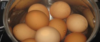
Nutritional value and nutritional value
The calorie content of a chicken egg is no more than 17%, due to which this product is considered one of the main ones when following a diet. The product contains many amino acids. Ten of them are irreplaceable - they are formed in the body, and they can only be obtained by consuming egg products.
Protein is a necessary component of the human body, because it can be broken down into important amino acids required for the normal functioning of not only muscles, but also the human brain. The yolk is a higher calorie component, including many fats and fatty acids.
The eggs are similar in structure, but usually differ in size. The white and yolk, as well as the shell of eggs, contain many useful substances. This product is useful for humans, and its unique structure provides reliable protection for the future chick.
1
0
Copy link
Cytoplasm.
Oocytes contain a large amount of cytoplasm, which has a complex structure. It contains many mitochondria necessary to provide the cell with energy; the membrane system of the endoplasmic reticulum and numerous ribosomes on which protein synthesis occurs; the Golgi complex and lysosomes - the enzymes of the latter carry out intracellular digestion and can even initiate the destruction of the egg.
Microtubules were also found in young insect oocytes, which apparently participate in the movement of the cytoplasm. They are rare in the eggs of other invertebrates and in vertebrates.
In addition to this set of organelles, which are also characteristic of other cells, the cytoplasm of the egg in many cases contains the so-called. cortical granules, or bodies, which in a number of animals play an important role in fertilization. However, its most important feature is the presence of yolk, which is necessary to nourish the embryo.
There are at least three possible ways to form the yolk. Firstly, it can be produced by oocyte organelles. Secondly, the yolk precursors, i.e. the substances from which it is formed can be produced not in the oocyte, but in other cells and enter the oocyte by endocytosis. Finally, a combination of these two processes is possible. see also
CELL.
Main functions
The main functions of the testicles are the production of testosterone and the formation of sperm. In medicine, there are two types of functions - endocrine and spermatogenesis. Testicles simultaneously perform the function of internal secretion organs - they form hormones and carry them into the blood.
Endocrine
The endocrine function of the testicles is intrasecretory; it consists in the formation of sex hormones: testosterone, androsterone, dihydrotestosterone, dehydroapiandrosterone, which enter directly into the blood. Androgens are synthesized by Leidig cells lying in the interstitium between the convoluted seminiferous tubules.
The endocrine function of the testicles is as follows:
- development of a male body type, for example, broad shoulders, a rough voice, physical endurance, strong muscles;
- formation of courageous character, activity, endurance, desire for leadership, strong muscles;
- regulates cholesterol levels;
- responsible for the normal level of sexual activity;
- development of internal and external genital organs;
- hair development.
Reference! The production of testosterone is influenced not only by the gonads, but also by a man’s lifestyle - his diet, weight, physical activity, health and environmental factors.
The endocrine function of the testicles precedes the function of spermatogenesis, since under the influence of sex hormones the formation of the functioning of the genital organs occurs.
Spermatogenesis
Spermatogenesis is an important process that is responsible for the function of childbirth and occurs in men throughout their lives. During puberty in boys, the process of development of germ cells occurs, which lasts until old age.
There are cases where hundred-year-old men became fathers.
Spermatogenesis consists of four periods:
- Reproduction (carried out in the prenatal period, represents the division of germ cells after birth and until puberty. Upon reaching sexual development, some of the cells continue the process of forming new cells, the second part moves closer to the center of the tubule).
- Maturation (biochemical processes that are involved in preparing cells for the formation of sperm).
- Formation (sperm produce special enzymes that dissolve the shell of the egg, which allows them to penetrate inside and carry out the fertilization process).
Spermatogenesis lasts 75 days. This process is very delicate at the biochemical level, overly sensitive to the effects of adverse factors, such as:
- increased body or air temperature;
- chronic stress;
- taking hormones, antibiotics and other medications;
- excessive drinking;
- smoking;
- sedentary lifestyle;
- prolonged abstinence or sexual abuse.
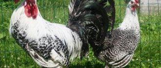
Are crosses better than the breed?
Starting from the second half of the current century, the process of rock formation has slowed down sharply. What is the reason for this phenomenon? It turned out that it is more profitable to create not breeds, but crosses, or rather, combining lines of chickens, when crossing which, due to the effect of heterosis (superiority of the offspring over the parental forms), the best productivity indicators are achieved.
Currently, the most complete gene map among farm animals has been compiled specifically for chickens. It has been determined, in particular, that the karyotype (a typical set of chromosomes for a species) of a chicken contains 78 chromosomes. What is a chromosome? This is one of the elements of the structure of the nucleus; the chromosome consists of DNA (deoxynucleic acid) and protein.
Each biological species is characterized by a certain number and shape of chromosomes. For geneticists and breeders, chickens are an excellent object for studying issues of heredity, since they have many known genes (more than 50 pairs have been discovered) that determine the formation of various qualitative traits, such as:
- ridge shape,
- coloring of feathers, body skin and legs,
- egg shell color,
- fledging speed.
Each of these traits is controlled by a known pair of genes, and it is not difficult to breed the desired individuals. The situation is more complicated with such economically important traits as egg production, weight and quality of eggs.
These traits are polygenic in nature and are determined by many genes. The total number of such genes and their location on chromosomes remain unknown to science. At the same time, it cannot be said that a modern genetic researcher or poultry breeder is wandering in the dark, groping for the desired genotypes.
The first stage in selection
On the one hand, the world experience of chicken breeding has given a lot, and, on the other hand, a lot becomes clear from indirect signs that are known from marker genes, or signal genes. Knowledge of the nature of inheritance of certain traits, their genetic and phenotypic conditionality determines the first stage of success in chicken breeding.
Second stage in selection
The second stage is associated with the creation of optimal conditions for keeping chickens and their adequate feeding. Only with this combination can more and more productive lines and crosses of poultry be developed.
In our strict rational age, there is almost no place left not only for fighting and decorative, but also for some meat-egg breeds of chickens. These breeds in industrial poultry farming cannot compete with specialized egg or meat breeds. Currently, only those breeds and populations that are distinguished by exceptionally high productivity are preserved and used in poultry farming.
Skin structure
The structure of the skin is simple on the one hand, and complex on the other. Depending on how deep you look. We will try to succinctly provide as much information as possible about the composition of human skin. Here's the diagram:
- Epidermis. This is the top layer that serves as a barrier against the penetration of water, bacteria, and also protects against other negative factors. In addition, it allows you to understand a lot about the state of internal organs. For example, with cirrhosis of the liver and hepatitis, the outer covers become yellow. Diagnostic criteria can include rashes, peeling and other damage. The quality of the epidermis directly depends on the health of the body. It consists of five layers:
- Basal. It is the lowest layer of the epidermis, which is in direct contact with the dermis. The tasks of the basal layer are simple - maintaining moisture (it contains up to 70% water), as well as cell regeneration.
- Grainy. It consists of small cells that fit very tightly to each other. The task of this layer is to secrete intercellular fat and moisturize the skin.
- Brilliant. It does not happen everywhere, but only on the feet and palms. The purpose of this layer is to prevent friction and wear of the cover. Considering that these are the parts that are most often exposed to adverse effects, without this layer we would tear our hands until they bleed if we wanted to pull ourselves up or do any work with our hands.
- Horny. Well, directly, the most famous layer of the epidermis, which performs many tasks, and above all, maintaining immunity. The stratum corneum protects the skin from external influences, such as temperature, bacteria or excess penetration of water from the outside.
Spiky. Its characteristic feature is cells that look like spikes (hence the name). The spinous layer produces keratin, which is necessary to give the cover strength and elasticity. This protein is one of the main building materials of this organ.
We described the structure of the skin only in general terms, because entire monographs have been written on this topic. You can dig endlessly, right down to the organelles inside cells and their chemical composition. Obviously this is unnecessary.
Reasons for downsizing
Small size of the tissues is a signal that pathological changes are occurring in the male body. The normal size is 4-6 cm in length and 2.5-3.5 cm in width. Size reduction in men occurs for the following reasons:
- influence of harmful factors during intrauterine development;
- pathologies of pregnancy, in particular from the fact that a woman uses different medications;
- infectious pathologies or autoimmune processes negatively affect the tissue (sometimes the human immune system is able to “eat” the tissues of its own body, including the testicles);
- injuries;
- harmful effects of ionizing radiation;
- cancer processes;
- Klinefelter or Shereshevsky-Turner syndrome (and other hereditary diseases);
- hypotrophy or atrophy of the testicles due to cryptorchidism (this often results in small genitals);
- inflammation of the testicles, varicocele and other diseases of the male genital area;
- uncontrolled use of anabolic steroids by athletes.
A true decrease in size indicates that the tissue is gradually atrophying. Accordingly, the production of sperm and testosterone decreases in them. These processes negatively affect fertility and male hormonal balance. The disease is diagnosed by palpation (palpation). The doctor makes conclusions about testicular disease only after a comprehensive examination.
The danger of severe atrophy is that it stops producing enough hormones, which negatively affects health. In some cases, this is a prerequisite for cancer.
Maturation.
An egg can leave the ovary at different stages of maturation; this means that its nucleus can be either diploid (in this case, the process of meiosis is completed during fertilization) or already haploid. Thus, in many worms and mollusks, as well as in a number of mammals (dogs, foxes, horses), meiosis is at the prophase stage at the time of fertilization, i.e. the egg still retains a large diploid nucleus (germinal vesicle). In other mollusks, such as the common mussel ( Mytilus edulis
), and in many insects the mature egg is in metaphase of the first mitotic division;
in most vertebrates - in metaphase of the second meiotic division; in coelenterates and sea urchins, meiosis in the mature egg is complete and the nucleus is haploid. A number of animals are difficult to classify into any of these four groups. For example, the eggs of the starfish Asterias
can, under some conditions, be fertilized at different times after they are laid, when they are at different stages of maturation.
Follicular cells.
In many organisms, the egg is surrounded by a layer of follicular cells, the cytoplasm of which contains organelles similar to the organelles of the oocyte. While the oocyte develops, the cytoplasm of the follicular cells forms projections that sometimes merge with the microvilli of the oocyte. The function of follicular cells remains unknown in many animals. However, in insects such as dragonflies and fruit flies, follicular cells produce material used to form a secondary shell around the egg.
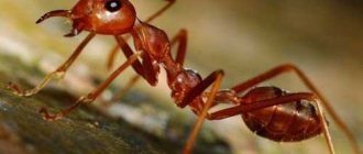
Trophic oocytes
There is a type of eggs that, when laid, serve as food for the offspring. As a rule, they are unfertilized, and their appearance is practically no different from ordinary ones. They are laid by the females of some ants and the queens of termites until the colony begins to produce enough food. In some cases, unfertilized oocytes of meat-egg and egg chicken breeds are also mistakenly called trophic, since they are used as food not by the birds themselves, but by humans and sometimes domestic animals.
PARTHENOGENESIS
Many invertebrates and lower vertebrates are characterized by parthenogenetic (virgin) reproduction, i.e. their eggs can develop without fertilization. ( See also
REPRODUCTION.) In some cases, for example in fish, this requires preliminary contact of eggs with sperm of individuals of another species: this activates the egg (inducing it to fragment), but does not fertilize.
A similar activation of eggs (both invertebrates and lower vertebrates) can be caused in laboratory conditions. To do this, they use methods such as pricking with a needle soaked in blood, keeping eggs at high or low temperatures, either in an acidic or alkaline environment, or in a hypertonic saline solution (i.e., in a solution with a higher concentration of salts than in the cage) , or in a solution of strychnine or saponin. If, as a result of such influences, it is possible to obtain a diploid organism, this usually occurs due to the suppression of one of the meiotic divisions or one of the first cleavages of the egg. However, with artificial parthenogenesis it is not always possible to achieve the full development of a new organism - most often the development of the embryo stops in the early stages. Therefore, in most cases it remains unclear whether these artificially induced processes correspond to normal development. It has been shown, however, that in the sea urchin Arbacia punctulata,
activation of eggs with a hypertonic solution, namely sea water with a high content of certain salts, induces processes similar to those observed during fertilization.
It was also possible to obtain complete and massive (from the vast majority of eggs) parthenogenetic development of the silkworm, using various physical (in particular, temperature) and chemical influences. It turned out that with a sufficiently strong effect on unfertilized eggs, meiotic division is inhibited in them, and in the future only females are hatched from such eggs. The same, but weaker effect, which does not inhibit meiosis but activates eggs, leads to the development of only males. Thus, with the help of artificial parthenogenesis, it is possible not only to cultivate this species, but also to regulate the sex ratio in the breeding population, which is important, since males produce more silk than females. This method of parthenogenetic breeding of silkworms has received practical application.
Interesting experiments were carried out on frogs. The nucleus of the frog egg was removed and the nucleus of a somatic cell was introduced instead. As already mentioned, the nuclei of all somatic cells, both embryonic and those taken from an adult organism, contain a diploid set of chromosomes, in contrast to the nucleus of haploid eggs. In a series of such experiments, oocytes of the clawed frog ( Xenopus laevis
) transferred diploid nuclei from blastula, gastrula cells, or from the brain of an adult.
It turned out that the cytoplasm of the oocyte is capable of changing the nature of the activity of the transplanted nucleus, regulating it in such a way that it corresponds to the activity of the cytoplasm. As a result, an oocyte with a transplanted diploid nucleus can develop into an adult frog. See also
CLONING.
About the mammalian egg, see
. HUMAN REPRODUCTION.