Published by: Parazitolog in Bacteria November 23, 2021
Malarial plasmodium: stages, types, development scheme Malarial plasmodium. Many insects are carriers of dangerous diseases. The malaria mosquito is perhaps the most famous representative of such insects. Malaria is a dangerous disease caused by Plasmodium falciparum. The infection occurs through the bite of a malaria mosquito, the saliva of which may contain specific enzymes and sporozoites of Plasmodium. As a result of a bite, a person experiences severe intoxication of the body, which is accompanied by severe fever. Malaria mosquitoes - carriers of malarial plasmodium. Only some mosquitoes of the genus Anopheles are capable of transmitting malaria. They are called malaria mosquitoes. This is a fairly extensive genus, including several hundred species. Not all representatives of the Anopheles genus are capable of transmitting malaria. Some are very good carriers, while others are not dangerous. These mosquitoes are widespread throughout the world. Including those...
Review
0
Evaluation criterion
Summary: Was the information useful? Please rate it
User rating 4.56 (1 votes)
Malarial plasmodium. Many insects are carriers of dangerous diseases. The malaria mosquito is perhaps the most famous representative of such insects. Malaria is a dangerous disease caused by Plasmodium falciparum. The infection occurs through the bite of a malaria mosquito, the saliva of which may contain specific enzymes and sporozoites of Plasmodium. As a result of a bite, a person experiences severe intoxication of the body, which is accompanied by severe fever.
- 1 Malaria mosquitoes are carriers of malarial plasmodia
- 2 Development cycle of malarial plasmodium
- 3 Periods and symptoms of the disease
- 4 Dimensions of malarial spasmodium 4.1 Sporogony
- 4.2 Tissue schizogony
- 4.3 Erythrocyte schizogony
Malaria mosquitoes - carriers of malarial plasmodium
Only some mosquitoes of the genus Anopheles
. They are called malaria mosquitoes. This is a fairly extensive genus, including several hundred species. Not all representatives of the Anopheles genus are capable of transmitting malaria. Some are very good carriers, while others are not dangerous.
These mosquitoes are widespread throughout the world. Including in those regions where malaria has been eliminated. They are also found in temperate climates; they are also found in the Moscow and Leningrad regions, Siberia, and the Far East.
Therefore, cases of local malaria are also possible in Russia. Since tourists who traveled to disadvantaged regions or migrants can serve as a source of mosquito infection. The risk is especially high in hot years. Mosquito larvae develop in water, so there are many more of them in damp places.
Contrary to popular belief, malaria mosquitoes are small (a few millimeters in length). The easiest way to distinguish them is by their characteristic stance, with the rear end of the body strongly raised upward (other mosquitoes keep their body parallel to the surface).
The presence or absence of the malaria pathogen in the mosquito’s body does not in any way affect its appearance or behavior. Therefore, it is impossible to distinguish between infected and uninfected mosquitoes without special analysis.
After the mosquito has drunk the blood of the patient, time must pass for the malarial plasmodium to go through certain stages of development and the mosquito to become infectious (usually 10–20 days). The rate of development of malarial plasmodia in the body of a mosquito is influenced by many factors, including temperature. At low temperatures, development slows down or stops.
Since the mosquito does not live long, it does not always have time to become infectious, dying before the malaria pathogen completes its development. This is one reason that indigenous malaria is rare in temperate climates. In addition, with the onset of cold weather, the circulation of the pathogen between people and mosquitoes stops.
Mosquitoes themselves do not contain malaria pathogens. To actually become malarial they must drink the blood of a person with malaria. A mosquito can only become infected from a person; malarial plasmodium is not transmitted to its offspring.
One of the main protective measures is to prevent bites from malaria mosquitoes. This is the use of nets and curtains (especially since most malaria mosquitoes prefer to feed at night), spraying insecticides indoors, choosing times and routes, and using repellents.
Plasmodium falciparum is the causative agent of malaria infection. According to the classification of protozoan (single-celled) parasites, they belong to the phylum Apicomplexa, class Sporozoa, order Haemosporida, family Plasmodiidae, genus Plasmodium.
There are 4 types of parasites that can parasitize the human body:
- Plasmodium vivax is the causative agent of tertian malaria;
- Plasmodium malariae is the causative agent of four-day malaria;
- Plasmodium falciparum is the causative agent of tropical malaria;
- Plasmodium ovale is the causative agent of ovale malaria, similar to the species P. vivax.
All varieties are generally similar in shape, structure and life cycles, except for details. But they have their own characteristics of the development cycle; mainly by the duration of its periods.
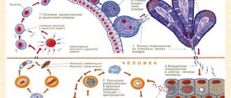
An interesting feature of parasites of the Apicomplexa type is the ability to alternate sexual (gamogony) and asexual (agamogony) methods of reproduction in the life cycle. In this case, a change of “master” occurs. In the human body, which is an “intermediate host” for the malarial plasmodium, only asexual reproduction is possible. And the phase of sexual reproduction is the prerogative of the main “host” and carrier of malaria, that is, the mosquito.
Infectious diseases.
Infectious diseases have occupied a major place in human pathology for many centuries and have been a scourge for the population. In pre-revolutionary Russia, millions of people fell ill with various infectious diseases every year; there was no year free from epidemics. Infectious diseases were the main cause of child mortality and general mortality of the population; they caused enormous damage to the population and the country's economy because they often left severe and irreversible consequences. Tens of thousands were blind after smallpox, deaf, deaf-mute after scarlet fever, and had persistent paresis and paralysis after meningitis and polio. The Soviet state faced a difficult situation with regard to infectious diseases from the very first days of its formation. It was further aggravated by the previous world war, the civil war that followed the revolution, foreign intervention, famine and devastation, and the huge migration of the population in the country. All this taken together led to the emergence of epidemics of unprecedented scale. Already in those days, doctors attached great importance to the fight against infectious diseases. Already among the first acts of the Soviet state were decrees and resolutions to combat these diseases. At the beginning of the 20th century, the fight against infectious diseases unfolded on a broad front. First of all, appropriate personnel were required. This problem was solved thanks to the organization of appropriate departments at medical institutes. No less importance was attached to the study of infectious diseases, which included specialists from various fields. Gradually, corresponding laboratories and institutes were created, in which (together with the departments of medical institutes) issues of infectious pathology were developed. Children's infectious diseases were isolated in a separate course because the most common infections, more often or almost exclusively, occur in childhood and even mainly in early or preschool age (they were even considered obligatory for children). For example, the incidence of measles was almost universal among children and was observed more often under the age of 5 years. Among those sick with whooping cough (the second most common childhood infection), half were children under 3 years of age. The incidence of chickenpox and scarlet fever is highest between the ages of 1 and 9–10 years. Infectious diseases took first place in the structure of child mortality, which was very high. Finally, all infectious diseases, including those that are common among both children and adults, such as dysentery, in children have a number of pathogenetic and clinical features due to the anatomical and physiological differences of the child’s body. In the 1920s, a consistently expanding study of infectious diseases in children began. It is necessary to note the leading importance of the work carried out by teams of scientists led by A. I. Dobrokhotova, M. G. Danilevich, A. A. Koltypin, V. I. Molchanov, and subsequently their students and followers. An exceptionally large contribution to the study of the pathogenesis of infectious diseases was made by pathomorphologists (V.D. Tsinzerling, A.A. Skvortsov, etc.). Various studies were carried out by domestic immunologists, microbiologists, virologists and other specialists; The most active work was carried out by epidemiologists. A huge amount of research has been carried out, unique literature has been created, and a coherent system for combating childhood infectious diseases has been developed, as a result of which unprecedented successes have been achieved in history. In our country, the incidence of morbidity has sharply decreased - even to the point of eliminating certain nosological forms; for all infectious diseases, the mortality rate has decreased, the mortality rate has decreased hundreds and tens of times; Childhood infections have almost lost their significance in population mortality. The sharply increased life expectancy in our country is generally recognized as the result of a decrease in child mortality and mainly due to childhood infections. A coherent system of combating infectious diseases has a state character. The work of healthcare workers, in particular their fight against infectious diseases, is an important part of the general creative activity of modern society. This fight is not over; the tasks of further neutralizing childhood infections remain to be solved. Currently, intensive development in the field of infectious diseases continues and leads to the discovery of new aspects of known diseases, the identification of new pathogens and even new nosological forms. Infectious diseases, under the influence of numerous factors, have undergone evolution, especially noticeable in recent decades. Therefore, the presentation of the course is characterized by increasing complexity and versatility when considering each nosological unit. A description of the classic manifestations of infections, characteristics of new forms of infectious diseases that have appeared in the process of evolution, the reason for their formation and, finally, modern features of pathogenesis and clinic are presented. Infectious diseases are diseases caused by microorganisms. They are distinguished by their contagiousness, the specificity of the pathogen, clinical patterns in the form of cyclicity and the formation of immunity during the disease process. Contagiousness consists in the transmission of the pathogen from the patient to surrounding people. The specificity of the pathogen is that this microorganism, when infecting other people, causes the same disease in them and this nosological form cannot be caused by other microorganisms. For example, typhoid fever, dysentery, diphtheria, and measles are caused, respectively, by typhoid bacilli, dysentery, and measles virus. Some other microorganisms may cause similar individual symptoms, syndromes, but not the entire complex of the disease. An infectious disease is often referred to as “infection.” However, these are not synonyms; the concept of “infection” is much broader and, along with infectious diseases, includes the so-called infectious inflammatory processes. The latter are also caused by microorganisms and are contagious, but do not have pronounced clinical patterns in the form of cyclicity divided into certain periods. The main difference is that the pathogen does not cause any specific clinical form, but a variety of diseases with their own clinical picture. Similar, up to complete clinical identity, diseases can be caused by other types of pathogens, for example, streptococcal, staphylococcal processes. Both streptococci and staphylococci have the ability to infect any tissue, any organ of the human body and cause various diseases: rhinitis, tonsillitis, otitis media, lymphadenitis, pneumonia, pleurisy, meningitis, sepsis, etc. Many of these diseases can be caused by other microorganisms. Thus, pneumonia can be caused not only by streptococci and staphylococci, but also by Pfeiffer’s bacillus, Klebsiella, etc. These infectious processes have not taken shape in the form of a specific infectious disease with its inherent cyclicity, their nosological characteristics are not sufficiently delineated. They usually occur in the form of inflammatory processes, their infectious nature is immutable. There are also differences regarding immunity. Infectious diseases provide predominantly strong immunity not only for years, but often for life (measles, chicken pox, mumps, whooping cough, typhoid fever, scarlet fever, etc.). Infectious processes either do not provide immunity at all or it is short-lived; in some cases there is even a tendency to recurrence of diseases, as is observed in relation to streptococcal and staphylococcal diseases. The division between infectious diseases and infectious processes is often very arbitrary; as we study, the lines between them become noticeably blurred. Individual infectious processes are united on the basis of common clinical, morphological and other signs, and most importantly - on the basis of the unity of the pathogen. Thus, streptococcal and staphylococcal infections were isolated and included in textbooks on infectious diseases. The main differences between infectious diseases are contagiousness, the specificity of the pathogen, the peculiarity of clinical patterns, and the formation of immunity. During infectious diseases, there is an incubation, or latent, period, a prodromal, or period of precursors, periods of disease development and convalescence. Infectious pathology must be understood in a broad sense, including all diseases caused by microorganisms - both infectious diseases and infectious processes.
Developmental cycle of malarial plasmodium
Malarial plasmodium. The development cycle of malarial plasmodium is quite complex. When a mosquito infected with malaria bites, sporozoites enter the human bloodstream (see below). In the human body, sporozoites primarily enter the cells of the reticuloendothelial system, forming primary tissue forms there.
This period of development of malarial plasmodium is asymptomatic. Subsequently, malarial plasmodia enter the blood and penetrate into red blood cells, completing a cycle of asexual development and reproduction there (schizogony).
The duration of one cycle of schizogony depends on the type of malarial plasmodium and determines the cyclicity of febrile attacks. Asexual forms of Plasmodium falciparum are called schizonts.
A mature schizont is divided into merozoites in the erythrocyte. After this, the erythrocyte is destroyed, and merozoites are released into the blood and then penetrate into new erythrocytes, undergoing a new development cycle there.
During tertian malaria, some of the merozoites can enter the cells of the reticuloendothelial system and form secondary tissue forms. The presence of secondary tissue forms determines the possibility of relapses of malaria.
Along with asexual forms (schizonts), sex cells can also be formed in erythrocytes - gametes: male (microgametes) and female (macrogametes). The presence of sexual forms of plasmodium in the blood does not give clinical manifestations, but is dangerous from an epidemiological point of view: such patients are infectious to mosquitoes.
Gametes, entering the mosquito’s body, undergo a sexual development cycle there, leading to the formation of a huge number of sporozoites that penetrate the salivary glands of the mosquito and are capable of infecting humans.
Life cycle of Plasmodium falciparum in humans: erythrocyte (clinical) stage of malaria
After the liver cells rupture, merozoites enter the blood and invade red blood cells. The erythrocyte (clinical) stage of schizogony begins.
Rice. 14. Erythrocyte schizogony. 5 and 6 are ring-shaped trophozoites. 7, 8 and 9 - young, immature and mature schizonts. 10 - erythrocyte merozoites.
Attachment to red blood cells
Attachment of merozoites to the membrane of erythrocytes and invagination into their membranes occurs due to the presence of special receptors on the surface of red blood cells. It is believed that the receptors on the surface of erythrocytes that serve as targets for merozoites are different for different types of Plasmodium.
Rice. 15. Red blood cells infected with Plasmodium vivax (the causative agent of tertian malaria) and Plasmodium ovale (the causative agent of tertian malaria) become enlarged, discolored and deformed, and toxic granularity appears in them. When infected with Plasmodium malariae (the causative agents of four-day malaria) and Plasmodium falciparum (the causative agents of tropical malaria), the shape and size of red blood cells do not change.
Erythrocyte schizogony
Having penetrated red blood cells, schizonts absorb the protein globin (a component of hemoglobin), grow and multiply.
In the erythrocyte, the parasite goes through 4 stages of development:
- Ring (trophozoid) stage.
- Stage of amoeboid schizont.
- Morula (fragmentation) stage. At this stage, the nuclei of schizonts are repeatedly divided (into 6 - 25 parts), and areas of the cytoplasm are separated around them. Erythrocyte merozoites are formed.
- Some parasites go through the stage of gametocyte formation.
A parasite that has penetrated an erythrocyte is called a trophozoid (schizont). It gradually increases in size. A vacuole appears near its nucleus and the trophozoid takes the form of a ring (ring) - a ring-shaped schizont.
As they grow (schizonts feed on hemoglobin), they increase in size and take on the appearance of an amoeba - an amoeba-shaped schizont.
Then, as the schizont grows, it becomes rounded and its nucleus divides many times—the morula stage. Each type of plasmodia has a certain number of nuclei: 12 - 12 in P. vivax, 6 - 12 in P. malariae and P. ovale, 12 - 24 in P. falciparum. This is how erythrocyte merozoites are formed. From the 1st schizont, 8–24 blood merozoites are formed, from which asexual and sexual forms of parasites further develop.
The duration of the erythrocyte schizogony phase is 72 hours in P. malariae, and 48 hours in other Plasmodium species.
During growth, pigment accumulates in the cytoplasm of parasites. Its appearance is associated with the processes of hemoglobin assimilation. Hemoglobin accumulations look like rods or grains. Its color ranges from golden yellow to dark brown.
Rice. 16. During growth, pigment accumulates in the cytoplasm of parasites.
After the destruction of erythrocytes, merozoites enter the blood, some of which re-enter the erythrocytes, others undergo a cycle of gametogony - transformation into immature germ cells, gamonts.
Together with merozoites, heme (the second component of hemoglobin) enters the blood. Heme is a powerful poison and causes acute attacks of malarial fever.
Cycles of erythrocyte schizogony are repeated every 3 days, in other types of malarial plasmodia - every 2 days.
Rice. 17. Destruction of the erythrocyte and release of merozoites into the blood.
Rice. 18. A merozoite of Plasmodium vivax (the causative agent of tertian malaria) that has penetrated into an erythrocyte (thin blood smear).
Rice. 19. Young trophozoids of Plasmodium falciparum (the causative agent of tropical malaria) under a microscope.
Rice. 20. Ring-shaped trophozoites of Plasmodium vivax, the causative agents of tertian malaria (ring stage).
Rice. 21. The photo shows an amoeboid schizont Plasmodium vivax (stage of an amoeboid schizont).
Rice. 22. The photo shows mature Plasmodium vivax schizonts (morula or fragmentation stage).
When red blood cells are destroyed and merozoites are released into the plasma, febrile attacks and anemia develop. When liver cells are destroyed, hepatitis develops.
Periods and symptoms of the disease
Malarial plasmodium. The entire process, from infection to recovery, can be divided into four periods: incubation, acute, latent and relapse. The nature of the course of the disease and the frequency of relapses largely depend on the type of malarial plasmodium - the causative agent. For example, the incubation period of the disease can vary from 7 days to 1 year, depending on the type of malaria infection.
General characteristic signs of the disease:
- elevated body temperature (39 – 40 degrees); chills and fever replace each other;
- “ache” throughout the body;
- prolonged headaches;
- gagging and diarrhea;
- profuse sweating after a hot flash;
- severe enlargement of the liver and spleen;
- pain in the liver area;
- general weakness and anemia;
- possible confusion;
- yellowing of the skin and sclera of the eyes;
- frequent and painful urination;
- loss of appetite;
- insomnia.
Individual signs and symptoms (for example: cardiac stabbing pain, asthma attacks, joint pain) are also possible and depend on the presence of “weak points” in the body and the functioning of the immune system. Sick children usually develop a rash on their body. Tremors of the limbs are also typical for infants.
As the underlying disease progresses, other pathologies may occur, such as cerebral vascular ischemia and necrobiosis in the kidneys. In case of complications of the disease, coma and rupture of the spleen are possible.
Treatment of malaria
The main goal of therapy for this disease is to prevent the occurrence/recurrence of attacks and completely destroy the pathogen. The disease malaria or swamp fever is more common in endemic areas, so travelers should take preventative measures in advance. Treatment of malaria is carried out with the help of drug therapy, as a rule, Primaquine, Chloroquine, Atabrine (quinacrine hydrochloride), Akriquine are used.
Medicines for malaria
Drug therapy for this disease is considered an effective method. There are proven medications for malaria that have been used for a long time. An example of such a medication is Quinine, which was replaced by Chloroquine for a while, but then began to be actively used again. The reason for this was the emergence and then spread in Asia and Africa of Plasmodium falciparum, which was resistant to Chloroquine.
Depending on the region where the infection occurred, certain drugs against Plasmodium falciparum can be used. Most of them are suitable for both treatment and prevention. Artemisia annua extract containing artemisinin and analogues of synthetic origin are highly effective, but also high in cost. The disease poses a great danger to residents who live in endemic areas where there is no access to drugs. In developed countries, there are no problems with purchasing medicines.
- Vegetable garden for the lazy
- Canned corn salad: simple recipes
- Pomelo fruit - benefits and harm, how to choose a fruit
Sizes of malarial spasmodium
The life cycle of human malaria pathogens consists of the following stages: the sexual process of reproduction in mosquitoes (sporogony); asexual reproduction in liver cells (tissue schizogony); asexual reproduction in erythrocytes (erythrocyte schizogony); formation of sexual forms in erythrocytes - gametocytes.
Sporogony
In the body of a nopheles patient with malaria or a parasite carrier, gametocytes enter the stomach of the insect: macrogametocytes (female) and microgametocytes (male). After restructuring of the nuclear apparatus, the macrogametocyte turns into a macrogamete. 4-8 microgametes are formed from a microgametocyte.
In the stomach of a mosquito, fertilization of a macrogamete by a microgamete occurs. As a result, a motile zygote is formed, called an ookinete. The latter penetrates the wall of the stomach and an oocyst forms on its outer side.
The nuclei in the oocyst divide repeatedly, after which sporozoites are formed inside the oocyst - elongated spindle-shaped bodies measuring 11-15 microns in length.
The oocyst shell ruptures and the sporozoites penetrate the salivary glands. When a mosquito bites, sporozoites enter the human body. The duration of sporogony depends on temperature. At temperatures below 16°, sporogony does not occur.
Tissue schizogony
Malarial plasmodium. Sporozoites can remain in human blood for no more than one hour. During this period, they penetrate the liver parenchyma cells, where schizonts are formed. Their development has been studied in detail on Plasmodium species parasitizing monkeys and partly on human Plasmodium.
The sporozoite, having penetrated the liver cell, becomes rounded, increases in size, and the nucleus of the resulting schizont is successively divided. By the 6-12th day, the parasite fills the entire liver cell, pushing the cell nucleus to the periphery.
The size of the parasite is up to 60 microns. Such a large schizont breaks up into a large number (thousands and tens of thousands) of merozoites.
These latter in P. falciparum penetrate erythrocytes and subsequently the parasites develop only in erythrocytes; in other species, merozoites penetrate erythrocytes, as well as into liver parenchyma cells, where they undergo subsequent cycles of tissue schizogony. The duration of tissue development in P. vivax vivax and P. ovale is 7-8 days, P. malariae 11-12 days.
Erythrocyte schizogony
Tissue merozoites penetrate the erythrocyte and form schizonts, which disintegrate into erythrocyte merozoites. Red blood cells are destroyed and the released merozoites settle in new red blood cells.
Malarial plasmodium. And some of the merozoites form gametocytes. The latter can circulate in the blood for a long time, their further development (sporogony) occurs in the carrier. Different stages of development of malaria pathogens in the blood are clearly distinguishable by morphological characteristics. However, P. vivax vivax cannot be distinguished from P. vivax hibernans.
The morphological characteristics of the pathogens themselves and the changes they cause in red blood cells make it possible to determine the type of parasite using preparations (smears and thick drops of blood). In the erythrocyte development cycle of Plasmodium, the following stages are distinguished: rings, schizont, merulation, young and formed gametocytes.
The size and shape of the parasite at different stages of development, the duration of the entire schizogony cycle, the number of merozoites formed and their size, the number of grains (lumps) of pigment, their color and location in the parasite, the shape, size and other characteristics of gametocytes, as well as changes observed in the infested erythrocytes, serve as signs to distinguish one species from another.
The development of the erythrocyte stages of all types of human malaria pathogens occurs in the circulating blood. An exception is P. falciparum, in which only ring stages and gametocytes are found in the blood; further development of schizonts up to the release of merozoites from erythrocytes occurs in the capillaries in which the deposited blood is located.
Complications of malaria
Timely provision of correct therapy ensures complete recovery in the vast majority of cases. Mortality under such conditions does not exceed 1% of the total. Lethal outcomes are caused not by the pathology itself, but by complications of malaria. Possible consequences of the disease:
- mental disorders;
- acute renal failure;
- cerebral edema;
- malarial coma (cerebral pathology).
Urgent timely therapy will help to avoid death and the development of complications. Kidney failure leads to an increase in nitrogenous waste in the blood, which will lead to infectious-toxic shock. The clinical picture of cerebral edema is usually observed in children with the fulminant form of malaria. Unlike adults, with the tropical form of pathology, a child may develop mental disorders. In case of death, the disease will develop in the following sequence:
- attack of fever;
- severe headache and cramps;
- there is a disruption in the functioning of the vascular and respiratory centers;
- respiratory and cardiac arrest;
- fatal outcome.
The structure of malarial plasmodium
Malarial plasmodium. Phylum Sporozoa: the phylum includes exclusively parasitic protozoa. In connection with the parasitic lifestyle, a simplification of the organization occurs (the disappearance of organelles for capturing and ingesting food, digestive and contractile vacuoles). The life cycle becomes more complex - a change of hosts, alternating asexual and sexual reproduction. The representative of the phylum is the malarial plasmodium.
Sporozoites are thin, worm-shaped cells that enter the liver cells through the bloodstream, where they transform into schizonts, which reproduce by multiple divisions - schizogony. In this case, the nucleus divides repeatedly, then a large number of daughter cells are formed from each cell.
The resulting merozoites leave the liver cells and invade the red blood cells. Here they feed, then schizogony occurs again. Thus, two forms of schizogony are distinguished - in liver cells and in erythrocytes.
Plasmodium falciparum causes malaria in humans. Infection occurs through the bite of a malarial mosquito (genus Anopheles), which contains the pathogen at the sporozoite stage.
As a result of erythrocyte schizogony, 10-20 merozoites are formed, which destroy the erythrocyte, enter the blood and infect subsequent erythrocytes. The cyclical nature of malaria attacks is due to the cyclical release of merozoites and their metabolic products from erythrocytes into the blood plasma.
After several cycles of schizogony, gamonts are formed in erythrocytes, which in the mosquito’s body will turn into macrogametes and microgametes. When gamonts enter the stomach of a mosquito, they turn into gametes, copulation occurs, the fusion of gametes. The zygote is mobile and is called an ookinete.
The ookinete migrates through the mosquito's stomach wall and develops into an oocyst. The oocyst nucleus divides many times, and the oocyst breaks up into a huge number of sporozoites - up to 10,000. This process is called sporogony. Sporozoites migrate to the salivary glands of the mosquito. Meiosis occurs after the formation of the zygote, sporozoites are haploid.
Thus, in the life cycle of Plasmodium falciparum, humans are the intermediate host (pre-erythrocytic schizogony, erythrocyte schizogony, onset of gametogony), and the malarial mosquito is the final host (completion of gametogony, fertilization and sporogony).
Schizogony
Malarial plasmodium. Plasmodium vivax causes three-day malaria, Plasmodium malariae - four-day malaria, Plasmodium falciparum - tropical, Plasmodium ovale - oval malaria, which is mainly characteristic of the inhabitants of Central Africa.
The French physiologist Charles Louis Alphonse Laveran discovered the human malarial plasmodium in 1890. Since 2004, another species has been identified - Plasmodium knowlesi, living in Southeast Asia. In addition, the latter type can cause malaria in long-tailed macaques (cynomolgus macaques, or cynomolgus macaques).
When bitten by the Anopheles mosquito, saliva enters the human bloodstream, containing specific enzymes that block blood clotting and Plasmodium sporozoites. Sporozoites are curved, fusiform, up to 15 µm long forms of plasmodium.
With the blood flow through the vessels, the sporozoites enter the human liver cells, where the asexual reproduction of the parasite begins. Schizogony of plasmodiums has distinctive features: the mother cell as a result of division produces not 2 daughter cells, as in other representatives, but many.
During tissue and erythrocyte schizogony, the formation of digestive vacuoles occurs in the protoplasm (internal contents of the cell) of merozoites. In these vacuoles, the nutrients necessary for plasmodium accumulate and waste products (toxins) that are not needed by plasmodium and are harmful to humans are removed from them.
Not all erythrocytes form new merozoites; in some, the formation of male and female germ cells - gamonts (hemotocytes) - occurs. Coming out of destroyed erythrocytes, merozoites penetrate into other (healthy) ones and divide again, soon destroying these erythrocytes as well.
Such repeated transitions occur with a constant frequency: in Plasmodium malariae every 72 hours, and in other species of Plasmodium - every 48 hours. With the same frequency, a patient with malaria experiences symptoms of intoxication (as toxins are released into the blood): chills and very high body temperature. These cycles of erythrocyte schizogony will be repeated until the number of merozoites necessary for further development is formed.
During tissue and erythrocyte schizogony, the formation of digestive vacuoles occurs in the protoplasm (internal contents of the cell) of merozoites. In these vacuoles, the nutrients necessary for plasmodium accumulate and waste products (toxins) that are not needed by plasmodium and harmful to humans are removed from them. Not all erythrocytes form new merozoites; in some, the formation of male and female germ cells - gamonts (hemotocytes) - occurs.
Sporogony
After several cycles of schizogony have occurred and gamonts have formed, the next stage of the plasmodium life cycle will begin. But for this it is necessary to make the transition into the body of the main host - the mosquito. This occurs when a mosquito bites an infected person. Together with the blood that drinks, gamonts enter the mosquito’s body.
Then, in the cavity of the mosquito’s stomach, the formation of mature germ cells - gametes - from gamonts and their fusion (fertilization) occurs. As a result of fertilization, a zygote is formed that penetrates the wall of the stomach.
Mature sporozoites from the wall of the mosquito's stomach pass into its salivary glands, and then, when a mosquito bites a person, they enter the human blood with saliva. And from this moment the entire life cycle of the malarial plasmodium begins anew.
Here the zygote becomes a growing and developing oocyst, which divides repeatedly to form thousands of new sporozoites. The process of formation of new sporozoites capable of reproduction through schizogony in the mosquito body lasts 7-45 days. This duration is influenced by the ambient temperature: the higher it is, the faster the sporogony stage.