Helminthiasis in pets is a completely common thing; all more or less experienced breeders and pet lovers have encountered them. But what association do you have from the word “worms”? You probably immediately remember the worms that live in the digestive tract of animals. Of course, this is not without reason: this is where the vast majority of parasites are localized. But don’t think that all worms are so “banal.” There are, for example, even subcutaneous worms in dogs!
What is Larva Migratory Syndrome?
The content of the article
Based on the developmental characteristics of the parasite, two types of hosts are distinguished: intermediate and final.
- The host containing a sexually mature worm is called final.
- The host containing the developing worm larva is intermediate.
Helminths, entering the host’s body, use its nutrients, are toxic to the nervous system, poison internal organs and therefore can cause various changes in the body.
Animal nematode larvae that enter the human body can cause larva migrans syndrome. Since humans are an intermediate host, pathogen larvae do not grow or mature in the human body, but can migrate through tissues and cause inflammation.
This syndrome can be caused by several types of helminths. However, it is most often caused by:
- round dog worms Toxocara canis;
- less commonly, cat roundworms Toxocara cati;
- or Toxocara mystax larvae.
The disease caused by Toxocara larvae entering a living organism is called toxocariasis. This is a dangerous chronic disease, it lasts a year or more with exacerbations. The prevalence of the infection is associated with the infection of dogs and cats and the contamination of the environment with their feces.
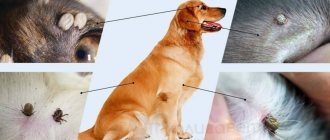
How to detect maggots?
Maggots are easy to spot; they actively scurry around in the animal’s fur. The nasty-looking parasites devour the rotting flesh of the victim alive, thereby causing unbearable pain to the dog. Infection with maggots is accompanied by the following symptoms:
- a pathia;
- aggression;
- severe itching;
- lethargic behavior;
- lack of appetite;
- intoxication of the body;
- heat.
Purulent wounds are a favorite place for maggots. They reproduce at maximum speed, since the dog’s body has all the comfortable conditions for their living: available food and warmth. In an open wound on a dog, you can easily notice elongated light-colored creatures with the naked eye. By feeding on the pet’s flesh, they will actively increase in size and absorb more and more soft tissue, which subsequently begins to decompose. Fly larvae, penetrating deep into wounds, eat through subcutaneous tunnels, causing even more harm to the dog. By eating away cells, maggots injure muscle and bone tissue. In addition, the spread of pathogenic microflora in the body further worsens. The skin tissue in the affected areas begins to swell and fester.
Toxocariasis - migration syndrome
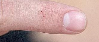
The larvae penetrate the skin. Toxocara eggs are found in large quantities in the soil. Toxocara larvae are most often found in children, affecting 2% to 10% of those infected. The incidence is the same in boys and girls. The age of sick children is from 1 to 7 years.
Infection of children with the disease is caused by:
- failure to comply with hygiene requirements;
- playing in the sandbox or with animals.
It has been established that in children aged 1–6 years, geophagia manifests itself as a behavioral disorder in 2–10%. This disorder is important because at the onset of intraocular larval migration syndrome, 40% of patients have this behavioral pathology.
The disease is less common among adults. In Western Europe, 2–5% are affected. Healthy adults have antibodies to Toxocara.
The incidence of toxocariasis also depends on the place of residence. Toxic pathogens are more common in tropical and temperate climates, and in warm countries their number reaches 86-93%. Infectiousness is much higher among rural populations (14-37%) compared to urban populations.
According to various countries, infection of Toxocara eggs in courtyards, sandboxes, beaches and other public places ranges from 10% to 30%. This problem is associated with an increase in the number of dogs and cats left unattended and non-compliance with the deworming schedule for pets.
Scabies in a dog: symptoms, how to treat and cure (what medicine to use)
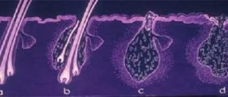
The causative agent of this disease is a tick no more than 0.25 mm long. Adult females penetrate the upper layers of the skin and lay 20 to 40 eggs there. In three months, each pair of ticks can produce offspring of up to 1.5 million.
Subcutaneous scabies affects animals of all ages. It is contagious not only to other pets, but also to humans.
Symptoms of mange in dogs occur with the appearance of small blisters on the bridge of the nose, around the eyes, at the base of the ears, or in other places. The liquid released from the bubbles dries in the form of scales similar to bran. They cause severe itching and hair loss in these areas, the skin becomes rough and wrinkled.
If left unchecked, the disease can cause gastrointestinal disorders and other illnesses, and after a few months even lead to the death of the animal. How to cure scabies in a dog is described further on this page, and several practical recommendations are presented. It is necessary to select the appropriate medicine for scabies in dogs with the help of a veterinarian who will make the correct diagnosis.
Subcutaneous scabies is very similar to dry eczema. It can also be confused with demodectic (red) scabies or ringworm. Subcutaneous scabies mites are much more difficult to detect than red scabies mites. To get to them, you need to consistently scrape the skin with a sharp blade. They can only be seen under a microscope or with a strong magnifying glass.
How to treat scabies in dogs with a guarantee of a high degree of effectiveness? Until a few years ago, scabies was treated with medications that required repeated applications. Today, dermatologists treat scabies with spregal. A bottle of medicine costs about $20. A single treatment of the skin of a sick animal is sufficient.
Pathogenesis
Pathogenesis
The final host of roundworms is a member of the dog and cat family, only in which the worm can grow, mature and lay eggs. When a worm enters a dog’s body, it parasitizes the intestines and feeds on the intestinal contents.
Dogs become infected with Toxocara by eating worm eggs in the soil. In the intestine, the larva released from the egg migrates with the blood to the lungs, bites the alveoli and enters the nasopharynx and mouth through the bronchi. This part of the migration, the airways, is called the tracheal stage of migration.
Later, the larvae enter the dog's intestines again through the esophagus, where they develop into adult worms that lay eggs. An adult female Toxocara produces about 200-250 thousand individuals. The remaining larvae penetrate the tissues and are distributed throughout the tissues of the body.
Studies have shown that in the female body, some of the larvae can remain in the tissues for a long time. In the last weeks of a dog's pregnancy, the larvae can become active and infect future puppies due to various hormones. Puppies can also become infected while feeding. Such an infected bitch can give birth to up to four puppies in a litter infected with Toxocara canis, without re-infection.
Thus, the main source of toxocara in humans is puppies under 3 months of age. Therefore, deworming pregnant bitches and all pets as needed is an important part of prevention.
With the dog's feces, the eggs fall into the soil and begin to mature, becoming an infection. Soil and related environmental conditions are important for egg development:
- air temperature (16-30 o C);
- air and soil humidity;
- light.
In soil, eggs mature in 2–3 weeks. Under the wrong conditions, the eggs go into a state of suspended animation and thus can survive the winter. Eggs can remain viable in soil for 8 years or more.
Humans are occasional intermediate hosts for this parasite and become infected with toxocariasis through oral feces. Unlike animals, larvae that do not enter the human body do not reproduce or grow, so people cannot become infected with them. Most often, the larva is transmitted through dirty hands when eating unwashed berries, fruits, and vegetables. It is also possible to become infected by eating contaminated and undercooked chicken, beef or lamb. Children can become infected while playing in the sandbox.
People cannot become infected with toxocariasis from a dog through contact, since the eggs must enter the soil and mature. However, care must be taken as dogs may carry mature eggs on their fur or tongue.
Mature Toxocara eggs enter the gastrointestinal tract through the mouth. In the human stomach, the eggshell dissolves, the larva is released and migrates towards the small intestine. It penetrates the intestinal mucosa and enters the blood and lymphatic vessels. Together with blood and lymph, the larvae enter the organs:
- liver;
- heart;
- lungs;
- kidneys;
- pancreas;
- eyes, etc.
It has been established that Toxocara cati larvae mainly migrate to the muscles, liver and lungs, while Toxocara canis larvae are often found in the central nervous system.
The larvae are encapsulated in tissues for a long period of time, after which the capsules disintegrate and the larvae are released. At this time, they become active and begin to migrate, irritating and damaging tissue. The movement of the larvae causes an immune tissue response and an allergic reaction.
This movement of larvae is called larval migratory syndrome. The condition can only be caused by the larval stage of the parasite and not by the adult worm or eggs. Depending on the location of the larvae, the syndrome is divided into two types:
- visceral;
- intraocular.
Weeping, wet and dry eczema in dogs: causes and photos of symptoms
Eczema in dogs, or summer scabies, is a fairly common skin disease among animals, including canines. In terms of symptoms, it is similar to mite scabies and ringworm. To exclude false diagnoses, you must first submit a sample for analysis.
Veterinarians are familiar with two forms of eczema: acute (moist, wet) and chronic (dry). Any of them usually appears in the summer.
Wet eczema in dogs, like all acute forms, develops quickly. Severe redness of the skin appears, the dog itches, and watery blisters form on the skin. Then the hair begins to fall out, the skin takes on a glossy, moist appearance. Most often, the acute form of eczema occurs in dogs of long-haired breeds.
Dry eczema in dogs does not make itself felt immediately, so if any inflammation, rash, causeless hair loss, or peeling accompanied by severe itching appears, you should immediately go to the doctor. This form of eczema most often affects wire-haired breeds.
Weeping eczema in dogs usually appears at the base of the tail, along the back and on the shoulder blades. The acute form is easily recognized by brownish discharge around the ear canal and between the toes.
Causes of eczema in dogs may include: local irritation of the dog's skin (from dirt or some chemicals), heat and poor diet, and nervous stress. It’s not for nothing that easily excitable dogs are more likely to get eczema, as are people with nervous disorders or metabolic disorders.
One very common type of eczema is caused by a microscopic fungus called Alternaria tenuis, which lives in dead grass and fallen leaves. Like most mushrooms, it loves moist, hot weather.
Eczema is not spread from a dog to another dog or person. If skin problems have spread to another dog, you need to urgently test for an infectious skin disease. The following shows eczema in dogs in the photo, which allows you to recognize the symptoms of the disease in an animal in time:
Before you begin to treat eczema, you need to remove skin irritants, wash your dog, and if the eczema is caused by lice or fleas, kill them. It often turns out that this is enough to combat eczema.
Treatment should be comprehensive and preferably early. First of all, it is necessary to act on the underlying cause within the body. Since the trophic function of the nervous system is always impaired in eczema, it is advisable to use short novocaine blockades or blockades of nodes and sympathetic ganglia at all stages. In the weeping stage, treatment of eczema is delayed, and it becomes chronic. After treatment, relapses are possible.
At all stages, for desensitization, it is advisable to administer a 10-20% solution of sodium thiosulfate intravenously, starting with 1-2 ml, adding 1 ml daily. A total of 10 injections are required.
To normalize trophic function and reduce exudation in the first stages before the formation of pustules, inject a 0.25% solution of novocaine with hydrocortisone (1 ml of novocaine solution 1 ml of hydrocortisone), and for more advanced forms it is recommended to add 1-2 ml of ampoule gentamicin. The solution is injected into several places on healthy skin so that it penetrates under the eczema-affected skin.
Clinic
Clinical symptoms of toxocariasis vary widely and depend on the organs and tissues affected by the migratory larvae. There may also be no symptoms.
The course of the disease depends on:
- number of larvae;
- affected organ;
- general resistance of the body;
- re-infection rates.
Since migratory larvae cause allergic inflammation, the severity of the disease also depends on the body's allergic reaction to the toxocarial larvae. In patients with atopic allergic disease, this infection is usually more severe.
Specific antibodies cause an inflammatory response to the toxocarsecretory (TES) antigen and the allergic component TBA-1. These antigens activate CD4+ cells and they differentiate into Th1 and Th2 cells. Interleukin-2 (IL-2) and γ-interferon (γIFN) secreted by Th1, interleukin-4 (IL-4) and interleukin-5 (IL-5) induce additional immune responses.
IL-4 promotes the proliferation and differentiation of B cells into plasma cells; when exposed to IL-2, IL-4, IL-5 and γIFN, they begin to synthesize antibodies of various isotypes. IL-5, in turn, promotes eosinophil differentiation and adhesion.
Epithelial inflammatory cells surround the larvae and form specific eosinophilic granulomas. It is the localization of granulomas that determines the clinical course of the disease.
There are different forms of visceral and ocular (intraocular) larva migrans syndrome. This classification is greatly simplified since one of these forms is always possible. And detection of toxocariasis of the eye and examination of internal organs usually reveal varying degrees of damage to internal organs.
Visceral migratory larval syndrome
Toxicosis can affect various organs:
- skin;
- lungs;
- liver;
- joints;
- heart;
- eyes;
- brain.
Thus, the syndrome manifests itself with various symptoms. However, the most common areas affected are the respiratory tract, making signs of upper respiratory tract inflammation one of the most common.
Unexpected manifestations of visceral larval migration syndrome are also possible after the sudden migration of a large number of larvae:
- acute pneumonia;
- eosinophilic meningoencephalitis.
This syndrome may increase the risk of death if the infection is accompanied by an abnormal immune response or when there is extensive migration of larvae into the myocardium or central nervous system.
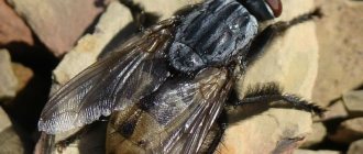
Development cycle
The female flies up to the future “incubator” and literally injects the larvae into the affected area of the skin.
Each “injection” contains at least 10-15 individuals. The larvae quickly burrow into the wound, actively feed and molt twice. After about five days (depending on the ambient temperature), they crawl out of their cozy nest, fall onto the soil, burrow into it, and pupate. After one to three weeks, the imago (that is, the adult stage) emerges.
Interesting!
Adult flies are most active at an ambient temperature of 20-27 degrees Celsius. If it gets hotter, they find shade and fall into a kind of torpor.
Intraocular larva migrans syndrome
This type of syndrome occurs when toxocariasis larvae enter the tissues of the eye and orbit through the central retinal artery and dentate arteries. Ocular toxocariasis is classified according to the 1971 Wilkinson and Welch classification according to the developing clinic. Only now the classification has become more precise as more is known about the syndromes.
Classification:
- Chronic endophthalmitis (vitreous rays, membranes, granulomas; posterior uveitis is pronounced; visual acuity is sharply reduced);
- Posterior granulomatous chorioretinitis (in the posterior pole of the eye, in the macula, on the optic nerve papilla or adjacent to these retinal structures there are 1-2 white lesions; usually visual acuity is reduced);
- Peripheral granulomatous chorioretinitis (white formations covered with vitreous connective tissue, penetrating the vitreous located on the periphery of the retina; visual acuity usually does not change).
In rare cases, ocular toxocariasis may manifest as:
- neuritis;
- neuroretinitis;
- localized vitreous abscess;
- iridocyclitis;
- larval keratoconjunctivitis.
If toxocariatic larvae enter the tissue in front of the orbital septum, this causes inflammation of the anterior septum cell: the eyelids and conjunctiva become swollen and red. When the larvae enter the orbital septum, the orbit becomes inflamed.
Cellular inflammation:
- swelling and redness of the joints and eyelids;
- painful eye movements;
- doubling;
- ophthalmoplegia;
- inversion;
- Optic neuropathy.
Most often, one eye is affected, and visual changes directly depend on the location of the lesion. Ocular toxocariasis is often complicated by:
- retinal circulatory disorders;
- retinal detachment;
- abscess of the eye;
- secondary glaucoma or cataract.
Diagnostics
Diagnosis
Diagnosis of all helminth infections is based on taking an accurate history. Epidemiological aspects are very important, since the risk of developing a particular infectious disease is closely related to:
- profession;
- rest;
- travel to certain endemic areas.
But they alone are not enough.
Of course, diagnosing toxocariasis is not easy, because people do not excrete toxocariasis worms or eggs, and it is also difficult to detect migrating larvae in biopsy material. Therefore, to confirm the diagnosis, instrumental and laboratory tests are always carried out:
- Complete blood count
: leukocytosis, persistent eosinophilia, anemia, thrombocytosis. Eosinophilia is reported to be more common in children than in adults; - Immunological tests.
More common: hypergammaglobulinemia. The level of IgE gamaglobulin increases. Increased isohemagglutinin titers for blood group A and B antigens. Antibodies to Toxocara are found in the vitreous body (90% of cases); - Linked immunosorbent assay
. Toxic secretory (TES) antigens are detected using direct ELISA. For toxocrosis, the specificity of the ELISA assay is 92–95% and the sensitivity is 73–75% when using a titer of 1:32 for diagnosis (4, 14). Indirect ELISA is used to detect antibodies (IgE, IgG and IgM) to TES toxocarial antigens. This method detects antibodies from 78 to 92%. cases; - Instrumental research methods
. In the visceral form of the syndrome, ultrasound or computed tomography reveals an enlarged liver, and intraocular examination reveals granulomas or abscesses in the tissues of the eye. Granulomas in the cortex or subcortex can also be seen on CT scans. - BMR Study.
Shows changes in the brain. Protein exudate in T1 and T2 modes may be more pronounced, and the potential difference may be visible. Gadolinium testing improves detection of abscess. Toxicara larvae can be identified by organ biopsy. It has been noted that larvae are extremely rare in biopsy material, but if they are not found, the diagnosis cannot be excluded; - Histological examination
. Multiple eosinophilic abscesses, allergic-type granulomas in damaged organs, and tissue necrosis around macrophages and lymphocytes were detected.
Cardiopulmonary form
Heartworms pose a serious threat to the health and life of a dog, as they affect vital organs. The longer they remain in the heart or lungs, the more damage heartworm disease causes in dogs.
Worms provoke dangerous pathologies of the heart and lungs, from which the entire pet’s body suffers. The dog suffers from shortness of breath, cannot run fast and enjoy life, it is weakened and lethargic, even if it is still very young.
If the disease is not treated in the early stages, another threat appears. When special drugs are used, the worms die, but their remains can enter distant areas of the lungs, causing thromboembolism.
A weakened or old animal may not survive such treatment, so in such dogs only methods of destroying the larval stage - microfilariae - are used in order to wait for the natural death of the adult helminth.
In this case, the dog must be provided with comprehensive symptomatic and supportive treatment. This does not always work, and if the infection is intense, it may not be possible to cure the dog - it will die.
Cardiac dirofilariasis in cats is less common, but cannot be ruled out. Most often it is found in stray animals living in basements, where mosquitoes that carry nematodes actively breed.
Pathogenesis of treatment
Detoxification therapy is used, including intravenous infusions of electrolytes. To restore dehydration, a 20% mannitol solution is administered intravenously. Intravenous or intramuscular administration of furosemide until the desired result is achieved. Nonsteroidal anti-inflammatory drugs (NSAIDs) are used to reduce the inflammatory response by reducing the activity of cyclooxygenase and the synthesis of prostaglandins and leukotrienes.
Damage to the central nervous system in larvae can lead to agitation and is suppressed by sedatives. For the form of ocular toxocariasis, all of the above drugs are prescribed. In severe cases or in the presence of complications, surgical treatment may be used:
- vitrectomy;
- cataract surgery;
- laser photocoagulation;
- enucleation of the eye.
Prevention
Toxicar eggs pollute the environment, so it is very important to follow the basic rules:
- personal hygiene;
- deworming of pets;
- clean environment;
- security of the children's playground.
Personal hygiene: every time you return from the field, you must wash your hands after stroking sand, soil and petting pets. Also, do not allow dogs to lick their faces or utensils used by people. Eat only washed berries, fruits, and vegetables.
Pets need to be dewormed promptly. Because puppies and kittens can become infected by transplantation or through breast milk. Deworming should begin after 3 weeks and continue every 2 to 4 weeks. Up to 6 months Puppies over 6 months of age are recommended to be dewormed every month. For adult dogs every 3 months. Bitches should be additionally dewormed 2 weeks before mating.
When going for a walk with your pet, be sure to pack bags to collect feces. This will prevent animal feces from polluting the environment. Pets should be avoided on playgrounds. When children are not playing, sandboxes should be covered. The sand in the boxes also needs to be changed regularly. The main and biggest problem is stray dogs and cats, so catching them and transporting them to shelters is an integral part of professional tactics.