The cattle skull is large and massive. It has the shape of a rectangle. The top side of the rectangle has a clear line. It is formed by horny processes and a bone ridge.
The bull's skull is distinguished by a stronger and wider horny ridge. It creates a reliable shield for the brain box. A cow's skull is more elongated. Their front part is elongated. The facial bone of an ox is shorter and stronger. The cranium connects to the cervical spine. The occipital part is massive and strong. It holds the neck muscles. What parts does a cow's skull consist of? What role do they play in the structure of the skeleton?
Cow skull, photo
Fig.1. Cow skull on the left side. Bone boundaries are shown.
1—nasal bone; 2— premaxillary bone; 3— lacrimal bone; 4—upper jaw; 3—zygomatic bone; 6—frontal bone; 7— parietal bone; 8—temporal bone scales; 8′—petrous bone; 9—occipital bone; 10—frontonasal suture; 11— lacrimal suture; 12— lacrimal suture; 13— lacrimal zygomatic suture; 14—incisor-maxillary suture; 15— lacrimal suture; 16— zygomaticomaxillary suture; 17, 18—frontal wedge-shaped suture; 19—sphenoparietal suture; 20—frontozygomatic suture; 21— lower jaw; 22—temporomygomatic suture; 23—frontoparietal suture; 24—temporoparietal suture; 25—occipital-mastoid suture.
Fig.2. Sagittal section of a cow's skull.
1—frontal bone; 2— parietal bone; 3—temporal bone (3a rocky part, 3b squamous part, 3c tympanic part); 4—occipital bone; 5—sphenoid bone; 6—ethmoid bone (6a dorsal concha, 6b perpendicular plate, 6c middle concha, 6d labyrinth of the ethmoid bone); 7—ventral shell; 8—nasal bone; 9— premaxillary bone; 10—upper jaw; 11— opener; 12— palatine bone; 13— pterygoid bone; 14— lower jaw; 15—mandibular foramen; 16—frontoparietal suture; 17—frontal wedge-shaped suture; 18—temporoparietal suture; 19—temporal wedge-shaped suture; 20—occipital-parietal suture; 21— occipital-mastoid suture; 22—occipital-sphenoid fusion; 23— pterygopalatine suture; 24—frontonasal suture; 25—transverse palatal suture; 26—incisor-nasal suture; 27—incisor-maxillary suture.
Fig.3. Cow skull from the dorsal surface.
1—frontal bone; 2— parietal bone; 3—zygomatic bone; 4— lacrimal bone; 5—nasal bone; 6—upper jaw; 7— premaxillary bone; 8—temporal bone; 9—frontoparietal suture; 10—frontal suture; 11— lacrimal suture; 12—frontonasal suture; 13— lacrimal zygomatic suture; 14—maxillozygomatic suture; 15— lacrimal suture; 16— lacrimal suture; 17—nasomaxillary suture; 18—incisor-maxillary suture.
Fig.4. Cow skull from the ventral surface.
1—occipital bone; 2—frontal bone; 2′—horny process; 3—temporal bone scales; 3′—tympanic part of the temporal bone; 4—body of the wedge-shaped host; 4′—temporal wing; 4″—orbital wing; 5— opener; 6— pterygoid bone; 7—zygomatic bone; 8—upper jaw; 8′—its palatine process; 9— palatine bone; 10— lacrimal bone; 11— premaxillary bone; 12—parietal bone; 13— occipital-mastoid suture; 14—occipital-sphenoid fusion; 15—sphenoid-temporal suture; 16—sphenoid-frontal suture; 17—temporomygomatic suture; 18— lacrimal suture; 19— zygomaticomaxillary suture; 20—transverse palatal suture; 21—median palatal suture; 22—occipital-parietal suture; 23—incisor-maxillary suture; 24—palatal incisal suture.
Udder structure
How many stomachs does a cow have: structural features of the digestive system
The udder is the part of the body for which the animal is so valued, therefore, when considering the anatomy of a cow, this organ should not be missed.
Circulation
Most of the udder is penetrated by a huge number of blood vessels. They saturate the organ with oxygen and take away metabolic products. During milk production, blood circulates from one part of the udder to another at a very high speed.
Lymphatic system
The lymphatic system in this part of the body is well developed. It controls the amount of fluid and removes accumulated toxins and impurities. Each part of the udder has its own lymph node, which swells if the animal has mastitis.
Thanks to the nervous system located in the udder, milk formation and instinctive milk production occur. The nerves send corresponding impulses to the brain, to which the animal reacts reflexively.
Milk follicles
Mammary follicle is part of the mammary gland. Its size and functions depend on the lactation period. It contains special ducts through which milk flows through channels and pipes.
The nipple is a muscular fold that has a sphincter at the end that closes the nipple canal. Thanks to it, milk does not spill out of the udder randomly.
Knowledge about the structure of a cow’s skeleton will allow us to understand the basic needs of a living organism, choose the right nutrition, and improve the living conditions of the animal.
Skeleton of the neck, torso and tail
How many bones does a cow have in total?
In cattle, the bone is compressed dorsoventrally and greatly expands caudally. The handle is massive, cylindrical, the anterior end is raised dorsally; connects to the body of the bone with a joint. The notch for the first pair of ribs is located at the front end of the handle.
Cervical region
In cattle, the vertebrae are massive and short; the heads and pits are well defined; the spinous processes are developed, have thickened ends, and their length increases in the caudal direction. The costal processes are located ventral to the transverse processes and are deflected forward. The ventral crest is present only on the 3rd - 5th vertebrae. On the 6th vertebra the costal process is wide and long.
Thoracic region
In cattle, the bodies of the thoracic vertebrae, due to their small number (13, rarely 14), are longer than wide. Wide, lamellar, with sharp caudal edges, the spinous processes are strongly inclined caudally. The longest process is on the 2nd vertebra. Diaphragmatic vertebra 13th. Sometimes the ends of the spinous processes are thickened, but flattened from front to back. There are lateral vertebral foramina. The costal facets on the transverse processes are saddle-shaped.
Lumbar
Cattle have 6 lumbar vertebrae; the cranial articular processes are equipped with grooved facets, the caudal ones are cylindrical. The vertebral bodies are long, with ventral ridges, narrowed in the middle. The transverse costal processes are set horizontally or even slightly bent dorsally at the ends; their edges are often jagged and sharp; the length of the processes of the 3rd and 4th vertebrae is greatest. The spinous processes are wide and low. The caudal intervertebral notches are deep.
Cattle have 5 vertebrae. The spinous processes are fused into a ridge with a thickened free edge. The wings of the sacrum are compressed from front to back; ear-shaped articular surfaces are directed caudally; cranial articular processes with grooved facets. The median vascular groove runs on the concave pelvic surface of the sacrum. The ventral sacral foramina are extensive. The fusion of the sacral vertebrae occurs at 3-3.5 years.
Tail section
Cattle have a long tail; there are only 18-20 vertebrae in it, but their bodies are significantly elongated in length. The rudiments of the arches are quite well defined. Of the articular processes, only the cranial ones have been preserved, and the transverse processes have taken the form of wide plates curved ventrally. There are hemal arches attached to the cranial ends of the bodies of the first 3-5 vertebrae, but more often there are only their rudiments in the form of tubercles on the first 9-10 vertebrae.
Components of the skeleton
The general features of the skeleton of animals related to cattle are also inherent in cows. There are differences between the sexes. Males have larger body parts and a more massive skeleton than females. In bulls, the frontal region is significantly prominent.
The skeleton of a cow consists of the bones of the skull, neck, torso, limbs and tail.
The skull is followed by:
- vertebrae of the cervical spine;
- thoracic vertebrae and ribs;
- shoulder blade;
- xiphoid cartilage;
- sternum;
- lumbar vertebrae;
- sacrum and ilium;
- Maklok;
- pubic and ischial bones;
- tail.
The forelimbs have bones:
- radial;
- elbow;
- wrist;
- metacarpus;
- sesamoid and fetlock;
- coronoid and hoof.
On the hind limbs are:
- femur and tibia;
- trochanter;
- knee;
- process of the fibula;
- tarsus, calcaneal tubercle and toe.
Skeletal growth
Mastitis in a cow
Newborn animals of different age maturity differ in the proportions of parts and areas of the body: in calves, compared to cows, the ratio of the length of the legs to the height of the chest is 25% greater, and the ratio of the diameter of the chest to the distance between the breasts is 11% greater. The ratio of the width of the frontal region to the length of the head is 8% greater than in cows.
Age of animals, months.
Live weight, kg Material from the site https://wikiwhat.ru
Absolute skeleton mass, kg
Relative skeletal mass, %
https://zoovet.info/vet-knigi/101-anatomiya-zhivotnykh/domashnie-zhivotnye/8212-sravnitelnaya-anatomiya-skeleta-golovy
https://oferme.ru/zhivotnye/korovy/stroenie-5259/
https://mnogo-krolikov.ru/korovy/opisanie-stroeniya-cherepa-korovy.html
https://wikiwhat.ru/%D0%A1%D0%BA%D0%B5%D0%BB%D0%B5%D1%82_%D0%9A%D0%A0%D0%A1
Structure of the cattle skull
In the cattle skull there are 2 sections: facial and brain. Marrow bones protect the animals' brains. The front part serves as protection for the organs of vision, hearing, smell and touch. How is the brain part of the skull formed?
- The occipital part of the skull is short and narrow. On the outside there are muscle tubercles. They are formed by small sphenoid and occipital bones. There are large holes on the sides of the back of the head. They have short jugular processes that are curved inward. On the lower part there are sublingual openings. They are elongated and have the shape of channels. On the inside there is space for the passage of the vein. The occipital region connects to the parietal part of the skull.
- Parietal department. In calves, this section occupies the main part of the skull. The frontal bones of young animals are not yet developed. With age, the crown recedes a little back, towards the occipital region. It is replaced by the frontal parts. The bones of the crown on the sides participate in the formation of the temporal foramina. The back wall is connected to the back of the head. The parietal plates protect the cerebellum. The skull of a bull has stronger bones than that of a cow.
- Sphenoid bone. The temporal parts of the skull and eye sockets are adjacent to it. The sphenoid bone contains canals for nerve trunks. The channels are connected to the opening of the eye sockets. Nerves approach the organs of vision through the hole.
- Frontal bones. In cattle they are well developed, forming the frontonasal and orbital-temporal parts. The nasal bone is thick. In the anterior part it extends to the eye sockets. The sinus is located in the nasal region. The orbital-temporal part is separated from the nasal frontal crest. It has a concave shape and goes into the processes of the cheekbones. The frontal part of the skull ends with horny processes. In bulls it is shorter than in cows.
- Temporal bones. They are connected to the parietal and occipital parts. On the bone is the zygomatic ridge, which connects the temple to the cheekbone. On its surface there is an articular tubercle. In the temporal part of the skull there are openings for the hearing organs. The narrow openings of the ear canals lead to the tympanic cavity.
- Lacrimal bones. They are connected to the frontal and zygomatic plates. A small gap separates them from the bow. The lacrimal bone contains a fossa for the lacrimal sac. Lacrimal canaliculi lead from the fossa to the eye sockets.
- The cheekbones of cattle are massive. They are connected to the temporal and frontal regions. The facial section forms the skeleton of the animal's muzzle.
- The jaws are dorsal. The lower jaws of animals are powerful and wide. On the far part there is a maxillary tubercle. The bones have a toothless edge. Cattle do not have alveoli for fangs. At the level of the 3rd molar, the facial tubercle is visible. He is performing. At the level of 1 tooth, the infraorbital foramen opens.
- Incisive bones. On top they are formed by plates and look like a roller. There are no alveoli for the development of incisor teeth.
More on the topic: How to slaughter a bull?
- The palatine bones of cattle are well developed. The hard palate occupies most of the oral cavity. The vertical palate is formed by the ethmoid bone.
- The nasal bones are curved and elongated. They form a ridge on the inner surface.
- Opener. The plate connects the nasal bone, jaw bone and frontal sinus. Bone has a complex structure. A cow's vomer is stronger than a horse's.
- The ventral jaw has 8 alveoli for incisor teeth.
- Hyoid bone. It has an anterior hyoid process and posterior branches that lead to the larynx.
Igor Nikolaev
auto RU
In a newborn calf, all parts of the skull are connected by cartilaginous layers. They make bones mobile. This makes calving easier for the cow. Cartilage in animals becomes stronger and transforms into harder compounds closer to 3 years. In adults, parts of the skull are connected by bone tissue.
What is the head made of?
Now we’ll find out what parts a cow’s head consists of and what differences they have.
Horns
Many cattle have horns. The only exceptions are specially bred breeds in which breeders have removed this trait. From an anatomical point of view, horns are hard bone formations that are located in the head area.
Their growth is directly related to metabolism and is manifested by the appearance of rings. Inadequate feeding of calves can lead to delayed horn growth.
Did you know? Newborn calves do not have horns, after a month you can feel small tubercles, and at 2 months of age the horns are already clearly visible.
Eyes
Cows have monocular vision due to the symmetrical placement of their eyes on the sides of their heads. The eyes are large and framed by rather long eyelashes, which grow not for beauty, but for orientation in space.
The eyes are located in bony sockets and have the following structure: round in shape, slightly convex on the outside and flattened on the inside.
Cattle eyes have the same connection to the brain as humans. They consist of 3 chambers (outer, middle and inner). Protection from mechanical impact is provided by eyelashes, eyebrows and lacrimal apparatus.
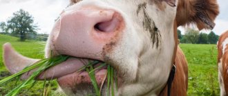
Teeth and tongue
The structure of cow teeth is not the same as that of humans. Incisors and anterior canines are present only on the lower jaw. The upper jaw is covered with durable keratinized epithelium. There are a total of 32 teeth in the mouth.
This jaw structure helps in tearing off strong grass. Retention of food occurs with the help of lips and tongue.
All ruminants swallow food without chewing. After some time, they regurgitate it, grind it with their molars and swallow it again. A newborn calf already has 20 baby teeth. After 1.5 years they are replaced by indigenous ones.
A cow's tongue is a collection of muscle fibers that give it the ability to move.
It performs the following functions:
- food tasting;
- help with swallowing;
- feeling objects;
- body skin care;
- contact with other animals.
Oral cavity
The oral cavity is a complex, well-coordinated organ responsible for the supply of food necessary for the normal functioning of the animal. It is here that food is crushed, moistened with saliva, and then sent down the throat.
Inside, the entire surface of the oral cavity (except for the teeth) is covered with mucous membrane. In some individuals, this shell has pigmentation of varying intensity. The organs of the oral cavity include: cheeks and lips, gums and teeth, hard and soft palate, salivary glands, tongue and tonsils.
The upper lip of the bull is connected to the nose and forms the nasolabial mirror. It can be used to judge the condition of the animal.
The cow's upper jaw remains motionless, while the lower jaw is able to make circular movements when chewing food.
Ears
The bovine hearing system consists of an outer, middle and inner ear. The outer ear detects sounds. It is represented by the auricle with fairly developed muscles, as well as the external auditory canal. The middle ear converts sound vibrations.
This is the eardrum with a chain of auditory bones. The middle ear has a connection to the pharynx. The inner ear is composed of bony and membranous labyrinths.
This is not only the organ of hearing, but also the vestibular apparatus of the animal. So, all the above information allows you to gain complete knowledge about the structural features of the cow’s skull and find out what parts it consists of.
Main anatomical features
The skull consists of 2 parts: the brain and the face. The first contains the brain, and the front part contains the cow's eyes, nose and mouth. In adult individuals, the facial part of the skull is larger in size than the brain part due to the developed jaws. And in small calves they are approximately equal.
How many teeth does a cow have? Calves have only 20 teeth. They are milky and by the age of 1.5 years they are replaced by permanent ones. An adult animal already has 32 teeth.
In a newborn calf, the bones of the skull are mobile relative to each other. Then, during growth, they are held together by bone sutures and form a strong connection.
The exceptions are the hyoid bone and the lower jaw.